Posts

Curves measuring less than 10 degrees: should we treat them?

As suggested by the Scoliosis Research Society (SRS), a scoliosis diagnosis is confirmed when a patient presents a Cobb angle measuring 10° or more and axial vertebral rotation. Maximum axial rotation is measured at the apical vertebra. (1) The SRS established this threshold in 1977, replacing the previous one of 7°. Ever since, 10 ° has conventionally been accepted, worldwide, as the threshold for diagnosing scoliosis.
However, structural scoliosis, with a potential for progression, can also be observed in the presence of Cobb angles measuring less than 10°. In fact, initial wedging of the vertebral bodies and disks can sometimes be registered with curves of 4°–7°. (2)

Idiopathic scoliosis, being a developmental disorder, most commonly arises and progresses during periods of accelerated growth (growth spurts).

The first such period occurs in infancy/early childhood, generally between 6 and 24 months of age, and the second between the ages of 5 and 8 years; finally, there is the pubertal growth spurt, which generally occurs at 11–14 years of age. (1)

Although the later stages of development are obviously not risk free, after puberty the rate of growth usually slows down, reducing the risk of progression of scoliosis. 

Can the risk of scoliosis progression be predicted in the case of curves measuring less than 10°?
There is, of course, always a chance that these curves will become more pronounced as the youngster grows, even, in some cases, to the point of requiring the use of a brace. But it is also true that most of them will remain stable over time without reaching the minimum criteria for a diagnosis of scoliosis. Certain factors may possibly be associated with an increased risk of scoliosis progression: a positive family history of scoliosis, laxity of ligaments, flattening of physiological thoracic kyphosis, a greater than 10° angle of trunk rotation (ATR), and growth spurts. All these factors should be evaluated by the attending physician. 

So, should we be treating these youngsters? In short, no. First of all, it is worth remembering, that the main aim of conservative treatment of scoliosis is to improve the patient’s appearance, but curves as mild as this rarely have an aesthetic impact; at most there may be some slight asymmetry of the trunk, but nothing that can be considered to exceed physiological parameters. With very rare exceptions, the only advice necessary in these cases is to opt for clinical monitoring of the patient, which can be considered to all intents and purposes a treatment, in the sense that it allows us to overcome the critical phases of development (which also correspond to the periods of greatest risk of progression of scoliosis) and also to intervene if any progression does occur. Monitoring is the first step in an active approach to idiopathic scoliosis, and it consists of clinical evaluations performed at regular intervals, ranging from every 2-3 months to every 36-60 months depending on the single case. 

In conclusion, any active treatment in this population of patients is actually overtreatment. Even just specific exercises, whose prescription constitutes first therapeutic step after monitoring alone, would cost these youngsters in time and effort, as well as being an economic cost.

A further aspect, not to be underestimated, is the psychological impact: starting a treatment amounts to confirming that the individual has a disease that needs to be treated, and this can lead them to start thinking of themselves as “sick”.

Furthermore, even though an exercise programme is not a particularly arduous undertaking, starting a treatment when there is no real need for one could compromise the youngster’s collaboration and commitment should a treatment be needed later on. This is an important consideration, because if their scoliosis does progress as they grow, specific exercises, rather than being useful, could become crucial, in order to avoid bracing for example.  

1 – 2016 SOSORT guidelines: orthopaedic and rehabilitation treatment of idiopathic scoliosis during growth

https://pubmed.ncbi.nlm.nih.gov/29435499/

2 – Radiographic Changes at the Coronal Plane in Early Scoliosis. Xiong, B., Sevastik, J. A., Hedlund, R., & Sevastik, B. (1994). Spine, 19(Supplement), 159–164. doi:10.1097/00007632-199401001-00008

https://pubmed.ncbi.nlm.nih.gov/8153824/

The experts’ brace-classification : a newly pusblished study with video-commentary

Studies have shown that brace treatment for AIS is effective but not all braces are the same. The classification of scoliosis braces developed by SOSORT with SRS, ISPO, and POSNA and approved by ESPRM, a study just published by The European Spine Journal, aims to produce a classification of the brace types.Studies have shown that brace treatment for AIS is effective but not all braces are the same. The classification of scoliosis braces developed by SOSORT with SRS, ISPO, and POSNA and approved by ESPRM, a study just published by The European Spine Journal, aims to produce a classification of the brace types.
Four scientific societies (SOSORT, SRS, ISPO, and POSNA) invited all their members to be part of the study. Six level 1 experts developed the initial classifications. At a consensus meeting with 26 other experts and societies’ officials, thematic analysis and general discussion allowed to define the classification (minimum 80% agreement).
The classification was applied to the braces published in the literature and officially approved by the 4 scientific societies and by ESPRM.
There are substantial differences in results published in the literature: one of the factors impairing research and leading to clinical confusion in the field is the absence of a classification to understand differences and commonalities among braces.
The only existing classification is common to all other orthoses, which is to classify braces according to the anatomical joints held underneath the brace—in the spine, these are the trunk regions. Unfortunately, according to this classification, almost all braces for spinal deformities fall in the thoracolumbosacral orthosis (TLSO) category, without other differentiations included.
As a result, clinicians cannot generalize research results on one brace to another with the same biomechanical action. Even worse, we could be inaccurately generalizing data on one brace to another brace with different biomechanical actions.
In this study the experts developed a definition for each item and were able to classify the 15 published braces into nine groups.
“This is the first edition of a brace classification that we expect to evolve further in future due to better understanding and more research – explains prof. Stefano Negrini, Scientific Director of Isico and first author of the article – It is based on expertise more than evidence, but we also must recognize that expertise is the first step of the pyramid of evidence when no better research data are available. Moreover, this expertise is shared worldwide among some of the best brace experts. The involvement and support of the leading scientific societies in the field should guarantee its dissemination”.
Watch the short video commentary of Prof. Stefano Negrini about the published study for our Isico Science corner video column

That hated hump!

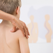
In a person with scoliosis, forward bending of the trunk will cause a protuberance to appear on their back, at the level of the scoliotic curve: this anomaly is commonly referred to as a hump.

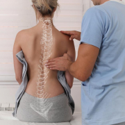
Scoliosis alters the alignment of the spine, and this alteration can be seen in the three dimensions of space: the affected vertebrae move sideways, taking on a different shape when viewed from the side; they also rotate, and this, especially if they are dorsal vertebrae, also causes a rotation of the rib cage. 

This hump is an aesthetic problem for people with scoliosis, as they fear that it is obvious to everyone else. But that is not the case at all. There is often a world of difference between the problem itself and how it appears in everyday life. 

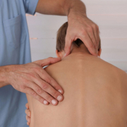
During a thorough postural analysis or a medical examination, the doctor or examiner will position the patient’s body to emphasise any asymmetries present precisely so that they can be identified and measured.

Nobody is perfectly symmetrical in daily life: when we move, we frequently twist our bodies and bend and rotate our joints, our movements involving different segments of our body in various combinations. As a result, we constantly develop asymmetries, humps, and combinations of bent and straight segments without even being aware of it.

Let’s take an example. A doctor examining a patient’s hump will have the youngster stand with his knees straight and body bent forwards so as to emphasise the protuberance on the side of the curve. Is it visible? Yes, if the patient’s scoliosis is sufficiently marked, it will be visible even to the untrained eye. 

Now, what if we ask someone with a healthy back to tie up their shoelace? To do this, most people will push their foot forward, bend their knees asymmetrically, and lean forwards, with their trunk deviating to one side (the side of the shoe needing to be tied). As they do this, their spine will be turned to one side, and a hump can be seen on their back, on the side of the leg, with the shoe needing to be tied. Does that hump mean they have scoliosis? Of course not! It is caused by them twisting their spine in order to reach their shoe.  

We can explain the scoliosis-induced asymmetry of the hips in a similar way. If we stand still with our feet parallel, our knees straight and our trunk aligned, our side and our hips will look symmetrical; in people with scoliosis, on the other hand, one hip will appear straighter in respect to the other. 

Now, let’s imagine how we normally stand. Do we ever actually stand with our feet positioned symmetrically and our bodyweight perfectly distributed between them? No! We usually stand with our weight on one foot and one hip thrust out. How do we look in this position? It is completely asymmetrical, but this asymmetrical appearance is due to our natural posture; it is not caused by scoliosis! 

Nobody ever notices these natural asymmetries because our bodies repeatedly assume them throughout the day. For this reason, even asymmetries caused by scoliosis are never really noticed by others. 

Going back to our “hump”: is it really such a bad thing, to the point that we should do everything in our power to get rid of it?

The back brace sometimes worn by youngsters with scoliosis, seeks to optimise the alignment of the vertebrae involved in the scoliosis curve, but how? It works by pushing “from the outside” directly on the affected portion to restore its symmetry. The force it exerts on the rib cage or on the soft tissues of the hips also affects the spine. 

 What is meant by an optimal alignment of the spine? The straightest possible? 

This isn’t an easy question to answer. Equally, it is not easy for the doctor to identify the correct balance of forces to be exerted on the spine by the brace pads. 

The best alignment will enable the treated spine to remain as stable as possible and to withstand the forces to which it is subjected in daily life. 

The strength of the spine depends to a large extent on its shape, as seen from the side. From this perspective, our spine is characterized by two curves, called lordosis and kyphosis. These curves must be well balanced: neither too pronounced nor too slight. 

Unfortunately, in scoliosis, and this applies particularly to dorsal scoliosis, the kyphotic curve is reduced, causing the back to appear flat or even hollow. In other words, the normal direction of spinal curvature is reversed, and the spine is weaker as a result. 

Unfortunately, the brace has no way of counteracting this problem, as all it can do is push. In fact, its corrective forces, which are applied to the hump to help align the vertebrae involved in the curve, have a flattening effect on the back’s upper part (dorsal section). 

Therefore, the doctor’s task is to find and maintain the best possible balance, considering the importance of all the planes of the spine.

What this means in practice is that it is sometimes necessary not to “go too far” in trying to eliminate the hump. Because if this objective can be achieved only by excessively reducing the dorsal kyphosis angle, the result will be a weaker and less healthy spine.  

Brace wearing: tenacity is rewarded with improvements

It is not just a question of how many hours the brace is worn but also patient compliance with the prescription. Take, for example, Marco and Ginevra. Both have idiopathic scoliosis, are followed by Isico specialists, and wear a Sforzesco brace for 20 and 23 hours a day, respectively. Marco complies with the prescription for wearing the brace, but he is inconsistent with the prescribed hours: sometimes he wears it for the 20 hours prescribed, others for 10, and others for 22. Ginevra instead regularly respects the prescription of 23 hours daily; this allows her to attain better results and avoid worsening.

Our study demonstrates this, Consistent and regular daily wearing improves bracing results: a case-control study published in the journal Scoliosis and Spinal Disorders some years ago. The study considered 168 patients who wore a brace for between 18 and 23 hours per day, divided according to high, medium, or low compliance and classified according to consistency or inconsistency in wearing.
The data were collected using Thermobrace, a temperature sensor applied to the brace to monitor its actual wearing.

Isico was the first organization to introduce Thermobrace into the daily clinical routine in 2010, and since then, its use has become commonplace. It has been verified that the relationship between doctor and patient is strengthened through Thermobrace, since the therapeutic choices are based on real data; therefore, the data obtained from the sensor can be used to facilitate the use of the brace.

“The data confirmed that the brace should be worn consistently, which means that wearing the brace for a constant number of hours allows the achievement of good results,” explains Dr Sabrina Donzelli, physiatrist and author of the publication, “also for those who are not completely compliant to the prescribed hours”.
This confirms what we always recommend to patients who undergo brace therapy: the break must always be constant; fewer hours one day and then recovering the lost hours in the following days is not ideal!

In addition to not adhering to the prescribed treatment, patients who are also not consistent in wearing the brace are at greater risk of worsening. Patients who have worn the brace for less than 70% of the prescribed duration are considered non-compliant.
“The study shows that to achieve the best results, the brace must be worn for a consistent number of hours. The attempt to recover lost hours is useless,” concludes Dr Donzelli “While tenacity together with compliance, i.e., the adherence of the patient to the prescription, is rewarded”. 

A judo champion in brace

My name is Giulia. I live in Genoa, and I am in my third year of Middle School. I am just like many other girls: I have my friends, I love the sea in summer, and I have my hobbies and interests, the main one being judo. 

In January 2020, I was diagnosed with idiopathic scoliosis. My parents requested several consultations before we ended up at ISICO.

It was a challenging and upsetting process for me and my parents, who struggled to know the best course of action to take.

To begin with, my treatment was just daily exercises: every month, I had a session with my physiotherapist, Martina, and every four months, a medical check-up with Dr Fabio Zaina, a physiatrist at ISICO.

It was quite a tough period because I had to do my exercises every day, regardless of homework, daily judo training, and holidays. When each check-up came around, I would get really anxious about what my curve would measure.

In April this year, it was found to have gotten worse, and Dr Zaina prescribed a brace. At first, I didn’t take it very well, as I started thinking about everything this would stop me from doing. I was thinking about my sport, my summer, going to the sea, and so on. My family were alarmed, too, but we soon calmed down thanks to Martina’s support.

It only seems like yesterday when Mum and I went to the orthopaedic office to collect my brace. I remember the tests and adjustments needed to make it feel as comfortable as possible. During the first “test run”, I really felt I couldn’t breathe and struggled to do even the most basic movements. Then, gradually, something seemed to change. Together with Mum, I started doing increasingly complex movements, like sitting down and getting up from benches in the play park close to where we live, walking faster and faster until I was doing short runs. By the end of that morning, it felt like my brace and I were getting to know each other: I was starting to adapt. The first night I managed to sleep quite well, and my new life began the following day. Luckily, my prescription was for 18 hours per day, so I could plan my days, concentrating all my sporting activities during my brace-off hours. 

In those early months, I started to measure this new situation, trying to set myself goals and then working to achieve them. I wanted to know how many things I could still do from my previous life with a brace on. 

So, I made a list and started to tick them off: walk to school with a backpack on; ride a bicycle; do a handstand (this one was pretty difficult); do a head-over-heels (I managed this after lots of tries). When the summer came, and I swapped sweatshirts for lighter clothes, I found myself having to try out new solutions and change my look a bit, opting for slightly looser T-shirts, but the brace wasn’t that obvious under them. And then, it was the school holidays.

Since I spend most of my summer on the beach, I had to rethink my brace-wearing schedule to fit some sun and sea into my brace-off hours and my training, which gets more intensive in the summer. To stick to the six hours allowed, I worked out a method that actually worked well: I decided that my brace-off hours would end exactly when my training sessions did and calculated them on that basis, counting back six hours to know exactly when they should start. At the allotted time, I would take off my brace and put on a costume to enjoy the sun and the waves. On the hottest days, though, even that solution seemed impossible, and after talking to Martina, I started removing the sensor from my brace so that I could also go in the sea with it on. Gradually, everything fell into place, and my everyday life became “easy” again, basically because I could still do pretty much everything I used to do. 

After a year’s break due to the pandemic, judo competitions started again. July brought the Italian Championships, my first ever. After taking part in, and winning, the regional qualifications, I went to Ostia for the national championships. I got through three matches and then won the final, which I was amazed about because I really hadn’t expected to. I automatically qualified for the next Italian Championships in November, thanks to that result.

The summer raced by, as there was so much going on – beach time, training, family holidays. Everything was just like normal!

In September, after returning to school, I started training for the new Italian Championships in November. I was to compete in the beginners’ B -40KG category: I was determined to defend the title I had won in the summer. So, I worked out a new daily plan, including school, gym sessions and free time, and threw myself headlong into this new challenge. 

On 14 November, I went back to Ostia, in some ways more excited than the first time but also more aware of the challenge I faced. Once again, I had four matches, and I retained my title as Italian champion in my category! I was even more thrilled than the first time because more athletes were competing on this occasion and, technically, the standard was higher. That was the day I realised that if you really want something and fight for it, nothing can stop you, even a problem like ours. We are just the same as everyone else!

When I first learned that I had to start wearing a brace, I read some of the stories of others like me fighting scoliosis. They said that your brace, in the end, becomes a kind of travelling companion, a friend who is always there for you throughout the day.  I didn’t really believe that, and I thought they were just words meant to encourage others like me.

But, you know, it really is true, and even though I would have preferred not to have to go through all this, it hasn’t stopped me from reaching the highest step on the podium and being happy!

Arabic version of the ISYQOL

Several questionnaires are available to assess Health-related quality of life (HRQOL) following conservative and surgical interventions for various spinal conditions.
Among these, the Italian Spine Youth Quality of Life (ISYQOL) questionnaire was developed to evaluate health-related quality of life in adolescents with Idiopathic Scoliosis and showed high validity in measuring HRQOL. 
After Chinese, Korean and Polish versions of the ISYQOL,  the study, Cross-cultural adaptation and validation of the Italian Spine Youth Quality of Life (ISYQOL) questionnaire’s Arabic versionaimed to undertake the process of adaptation of the questionnaire into the Arabic language (ISYQOL-Ar) and evaluate its validity and reliability, and to correlate it with the validated revised Scoliosis Research Society (SRS-22r)’s Arabic questionnaire in a cross-sectional multicenter study.
The ISYQOL was translated, back-translated, and reviewed by an expert committee. Reliability assessment for the questionnaire domains was performed using Cronbach’s alpha.
 A total of 115 patients were enrolled in the study and completed the ISYQOL-Ar and Arabic SRS-22r questionnaires.
The ISYQOL-Ar questionnaire is a reliable and valid outcome measure for assessing young patients with spinal deformity among the Arabic-speaking population.

Scoliosis in the mirror: aesthetic concerns and psychological support

We are all critical when we look at ourselves in the mirror, and we tend to focus on and analyse the smallest detail, to the point that we sometimes get quite worked up about perceived flaws or defects that others probably don’t even notice.
This also applies to curves and asymmetries due to conditions like scoliosis. Indeed, to a young scoliosis patient these changes can appear extremely prominent, even when their friends and relatives insist that they can’t see anything wrong with the youngster’s back.

First of all, a brief preliminary remark. As part of scoliosis treatment, it is crucial to consider the appearance of the patient’s back.
This is not only because improving its overall aesthetics is one of the aims of the treatment (Negrini et al., 2018), but also because any change in its appearance, major or minor, can have a considerable psychological impact on the individual in question.

Of course, the way we see our own body and other people’s bodies is highly subjective, and aspects of our own body that bother us might not be the same aspects other people notice.

“For some scoliosis patients, the presence of asymmetries of the waist or shoulder blades, or a hump, will be an unbearable problem, whereas other patients may be quite unconcerned about them” says Dr Irene Ferrario, Isico psychologist. “And this can be entirely independent of objective clinical assessments; it may depend on the individual’s mood at the time, or on how much importance is attached to physical appearance in a given setting”. 

Sometimes, patients’ concerns about their appearance become disproportionate, turning into actual fixations that lead to them pouring far too much time and mental energy into working out how they can conceal their perceived defects.

“Having said that, minimising the patient’s concerns or trying to convince them that they don’t have any defects can, in some cases, be counterproductive” warns Ferrario. “Indeed, in the most severe cases, trying to appeal to reason to dispel concerns only has the effect of losing you the patient’s trust.”

Why seek psychological support

Therefore, it is advisable to seek help from a psychologist or psychotherapist in these cases. Unfortunately, many patients are reluctant to take this step as they remain convinced that removing the physical defect is the only way to resolve the problem and feel better. 

This is why scoliosis treatment must also address psychological needs to be effective.

On the one hand, specialists need to understand their patients’ distress and show them that they recognise the reasons for it. Once it is clear that a patient feels heard and understood, the therapist should then gradually and cautiously begin the process of formulating and presenting other possible reasons for the distress, helping the patient to know where it comes from and how it can be impacted by their environment and the people around them.   

“When we look at ourselves in a photo or in the mirror, we think we are seeing exactly what other people see when they look at us,” says Dr Ferrario, “but our eye is well trained to focus on the things we do not like about ourselves and thus to play on our insecurities. Whereas others see us as a whole and do not to dwell so much on specific details, we tend to analyse ourselves in minute detail, and our attention is always immediately drawn to the particular thing we are uncomfortable with.”

The main objective of psychological counselling, therefore, is to reduce, as much as possible, the discrepancy between the way we would like others to see us (our ideal self), how we think others should see us (self-guide), and how others see us (our real self).

“The course of treatment we do with our patients aims to help them rebuild their body image by encouraging greater awareness of their strengths and weaknesses” Dr Ferrario concludes. “Gradually, over time, patients manage to internalise this new image and eventually form a more realistic view of themselves. Patients also become more accepting of the aspects of their body that they used to see in a negative light”.

The importance of observational studies

Our study, Observational Studies: Specific Considerations for the Physical and Rehabilitation Medicine Physicianwas recently published in the American Journal of Physical Medicine & Rehabilitation

Experimental and non-experimental designs are used to investigate the effect or association of an intervention and clinical or surrogate outcome. These methods aim to improve knowledge and develop new strategies to manage a disease or condition. 

While experimental research studies entail scrutiny by the scientist and provide results that are less prone to systematic errors, their downside is that they are poorly generalisable. “What all this means in clinical terms,” explains Dr Sabrina Donzelli, physiatrist at Isico and first author of the published research,” is that a treatment that worked fine during a study may in the long term, following its prescription by a hospital or general practitioner, throw up problems that did not emerge in the experimental research”. 

Therefore, to verify what happens in the real world, non-experimental studies, called observational studies, can be carried out, of the kind dealt with by the research we have just published. 

Well-designed observational studies can provide valuable information regarding exposure factors and the event under investigation

“Basically, what the researcher does is simply observe data, without having the possibility to manipulate it”, Dr Donzelli goes on. “The researcher’s task is to interpret and contextualise the results, taking into account all potential errors introduced during the selection of the study sample. To eliminate, as far as possible, systematic errors that could lead to incorrect evaluations and interpretations, it is necessary to implement a series of methodological strategies that are not very widespread in the rehabilitation field.” 

In physical and rehabilitation medicine, where complex procedures and multiple risk factors can be involved in the same disease, the use of observational study must be planned in detail and a priori to avoid overestimations. 

“This is why we wrote this article, to offer clear suggestions to researchers in the rehabilitation field who are interested in planning an observational study”, concludes Dr Donzelli. “We give an overview of the methods used for observational design studies and describe when it is appropriate to use them and how to do so in different scenarios”. 

The Risser sign, growth and scoliosis: let’s clear a few things up

When patients come for medical consultations or physiotherapy sessions, numerous measurements get taken and recorded, often without less expert eyes even noticing.

On the other hand, other measurements are quickly seized upon, both by parents and youngsters. Take height, for example. The sliding piece barely has time to touch the patient’s head before the patient, hopeful, blurts out: “Have I grown? Can I leave off my brace now?”

Another milestone we are promptly informed of is menarche in girls, as parents are often convinced that when their daughters start their periods, they have finished growing, meaning that their treatment can come to an end. But this isn’t always the case. On the contrary, this delicate phase can sometimes coincide with the most marked progression of the disease, making it all the more important to act with caution.

Although these are two important examples of the many factors that need to be taken into account to work out what point a youngster’s growth has reached, it has been shown that increases in height and menarche do not necessarily coincide with the individual patient’s growth peak [1] and may therefore not be helpful and/or sufficient when it comes to deciding on the best course of treatment.

Since these manifestations are secondary growth characteristics, they can only be seen as an indication that the patient’s growth spurt has begun. What they do not tell us is precisely how far on it is. There is a scientific explanation for the traditionally held belief that girls “develop earlier” than boys. In fact, because testosterone starts to be released into the body after oestrogens, boys start their pubertal growth spurt later than girls.[1]

To manage scoliosis and optimise the treatment results of the condition, it is crucial to have a good idea of the patient’s residual growth potential and the time remaining until he/she reaches skeletal maturity. An accurate prediction of the growth rate is also required to know when the deformity is likely to be most at risk of progressing. On the other hand, once it has been established with certainty that the patient has finished growing, this is the time at which preventive measures can be stopped with only minimal risk of further deterioration of the curve. [1]

There are various methods we can use to evaluate bone growth in adolescence, and one of them is called the Risser sign.

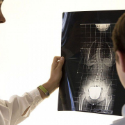
An individual’s Risser grade can be determined from an anteroposterior X-ray of the spine. An advantage of this method is that the same X-ray can be used to measure both the number of Cobb degrees (necessary to diagnose scoliosis) and the degree of skeletal maturity, thereby limiting the patient’s radiation exposure.

From 0 to 5, Risser grades are assigned based on the amount of calcification present in the iliac apophysis, and the scale thus measures progressive ossification. A Risser grade 0 indicates a low degree of bone maturity: this status is present from birth through puberty.

A Risser grade 5 means that the iliac apophysis has fused to the iliac crest, and the structure is 100% ossified: this status is present in adults  [2]. 

It would be misleading to imagine the transition from Risser 0 to Risser 5 as a continuous and constant progression that occurs over a fixed time and at a set pace. This is because growth is not constant but proceeds at different rates in the different phases. There are times when it pauses, times when it speeds up considerably, and times when it slows down.

The crucial stage in a youngster’s growth, also vital for understanding the course of their scoliosis, is the pubertal growth spurt, during which the disease can alter the shape of the patient’s back in the space of just a few weeks. From the perspective of a Risser evaluation of skeletal maturity, this stage corresponds to the transition from Risser 0 to the complete acquisition of Risser 1.

Between Risser 2 skeletal maturity and the end of the Risser 3 stage, the growth spurt slows down, but as far as the scoliosis treatment is concerned, we still cannot lower our guard: the patient should continue to receive treatment.

Scoliosis treatment is brought to an end gradually as skeletal maturity increases. Once the patient has reached Risser grade 5 (complete skeletal maturity), the treatment can be terminated safely without fearing that some of the hard-won gains might be lost 

The Risser classification varies slightly in different parts of the world, with some differences found, in particular, between Europe and America. In Europe, the successive grades tend to be assigned more cautiously, in the sense that a patient is deemed to have passed from one stage to the next only in the presence of precise levels of bone maturation. On the other hand, the American tendency is to assign the successive grades sooner.

Another method for assessing skeletal maturity is the Sanders classification, whose eight grades are assigned based on the assessment of hand bone growth [3]. Some studies have found the Sanders classification more precise than the Risser sign. It shows higher staging sensitivity when growth is most rapid and is therefore more reliable during certain growth phases [4]. The problem with the Sanders classification is that it requires a separate X-ray of the hand, which therefore means that it could increase the patient’s radiation exposure.

All this information clearly shows that residual growth is essential to evaluate, but at the same time, difficult to establish and interpret.

Specialists can, of course, use the classification they prefer, which will be the one that, in their experience, works best for identifying and evaluating the growth peak in adolescence. It is essential that they can correctly interpret all the data they collect, including from radiographs and patients themselves, to optimise the timing and results of the treatment. 

References

[1] Cheung JPY, Luk KD. Managing the Pediatric Spine: Growth Assessment. Asian Spine J. 2017 Oct;11(5):804-816. doi: 10.4184/asj.2017.11.5.804. Epub 2017 Oct 11. PMID: 29093792; PMCID: PMC5662865.

[2] Greiner KA. Adolescent idiopathic scoliosis: radiologic decision-making. Am Fam Physician. 2002 May 1;65(9):1817-22. PMID: 12018804.

[3] Sanders JO, Khoury JG, Kishan S, Browne RH, Mooney JF 3rd, Arnold KD, McConnell SJ, Bauman JA, Finegold DN. Predicting scoliosis progression from skeletal maturity: a simplified classification during adolescence. J Bone Joint Surg Am. 2008 Mar;90(3):540-53. doi: 10.2106/JBJS.G.00004. PMID: 18310704.

[4] Minkara A, Bainton N, Tanaka M, Kung J, DeAllie C, Khaleel A, Matsumoto H, Vitale M, Roye B. High Risk of Mismatch Between Sanders and Risser Staging in Adolescent Idiopathic Scoliosis: Are We Guiding Treatment Using the Wrong Classification? J Pediatr Orthop. 2020 Feb;40(2):60-64. doi: 10.1097/BPO.0000000000001135. PMID: 31923164.

Isico science corner video column

At Isico we give great importance to research, an integral part of our clinical work.  
Our newsletters always dedicate a space to the presentation and a comment when a new Isico study is published. 
From this issue on, we will go one step further, enriching each presentation with the video commentary of the first author of the research paper. We are pleased that it is just our scientific director Stefano Negrini, the first author of the article, launching the new Isico Science corner video column with a study of such great importance.