Do poor postures cause scoliosis?
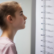
Parents often ask us: “My child often stands crooked or sits with a curved back – could this lead to scoliosis?”
According to the most recent scientific research, the answer is no: scoliosis does not develop as a result of poor posture. Several scientific studies have observed that some young people with scoliosis tend to adopt particular postures, such as standing with the pelvis slightly tilted or sitting asymmetrically (for example, with one leg tucked under the body).
However, it is important to clarify a key point: these postures are not the cause of scoliosis, but are very likely a consequence of a spinal curve that is already developing.
Recent studies, including those published in Scientific Reports (Yang et al., 2022) and BMC Musculoskeletal Disorders (Chen et al., 2024), confirm that certain postural habits or asymmetrical sitting positions are more common in adolescents with scoliosis, but they do not cause the condition. At most, they may represent the body’s adaptation to an existing spinal deformity or, in some cases, a factor that could slightly influence the progression of a curve that is already present.
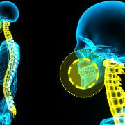
Idiopathic scoliosis, which develops during growth without a clearly identifiable cause, is considered a multifactorial condition, involving several elements, including:
- Genetic predisposition
- Rapid growth
- Hormonal and neuromuscular factors
- Mechanisms related to postural control
Posture, therefore, is not the origin of scoliosis, but may play only a secondary role within an already established condition.
That said, posture still matters.
While poor postures do not cause scoliosis, they may contribute to worsening an existing imbalance or making an already present curve more noticeable. For this reason, it is important to help young people maintain healthy postural habits: encouraging regular movement, avoiding long periods in the same sitting position, and paying attention to ergonomic conditions at school and during study.
Backpack use also deserves attention. School bags should not be excessively heavy and should always be worn on both shoulders, in order to avoid uneven loading. However, just like posture, backpacks do not cause scoliosis.
In summary
- Poor postures do not cause scoliosis
- They may be a consequence of the spinal curve or make it more noticeable
- Idiopathic scoliosis has complex and multifactorial causes
- Promoting good postural habits is beneficial for overall spinal health, but it is neither a treatment nor a prevention for scoliosis
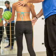
For this reason, whenever there is doubt, the most important step is an early specialist assessment, which can distinguish between simple poor posture and true scoliosis and, if necessary, guide the most appropriate management pathway.
References
- Yang Z, et al. Postural habits and lifestyle factors associated with adolescent idiopathic scoliosis. Scientific Reports. 2022;12:19203. PMID: 36344864
- Chen Y, et al. Association between incorrect postures and curve in adolescents with idiopathic scoliosis. BMC Musculoskeletal Disorders. 2024. PMID: 38744512