THE ROLE OF 3D ULTRASOUND IMAGING IN THE CLINICAL DIAGNOSIS AND MONITORING OF SCOLIOSIS DURING GROWTH
According to current guidelines, standing anteroposterior radiographs are the most reliable method for diagnosing scoliosis.
However, monitoring scoliosis, especially during growth, entails frequent radiation exposure (a full-spine X-ray every 6-12 months).
Even though there exists a low-dose X-ray system (EOS Imaging System), which we use with our patients, experts in recent years have been focusing on the quest to find an alternative, completely radiation-free system able to provide clinicians with equally reliable information.
Scolioscan®, developed in Hong Kong, is the first ultrasound imaging system that seems to offer valuable support in the clinical setting, both for monitoring scoliosis patients for progression of their curves and for screening populations for scoliosis.
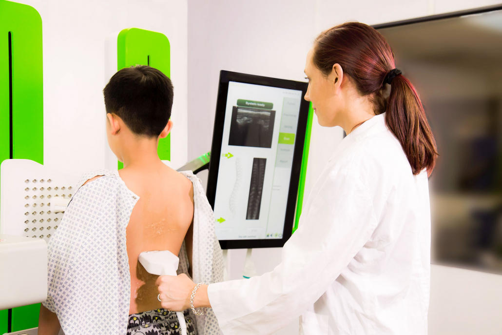
Let’s take a closer look at it. Scolioscan® is a 3D ultrasound system that generates spine images using a volume projection imaging method. For this purpose, the patient’s back is scanned using a linear probe (10 cm wide, with an ultrasound frequency of 7.5 MHz) equipped with an electromagnetic spatial sensing device that allows three-dimensional spine reconstruction.
The operator performs the scan freehand, using anatomical landmarks for reference: the scan starts from the sacrum (S1) and ends at the last cervical vertebra (C7).
It takes 30-60 seconds to perform, during which the patient has to be in a standing position, resting on four supports. At the end of the scan, the software immediately produces 9 images corresponding to progressively deeper coronal sections. The operator can thus select the image that gives the best view of the spinous and transverse processes of the vertebrae.
Thanks to these landmarks, the software reconstructs the curves based on the ultrasound spinous process angle (USSPA) or the ultrasound transverse process (i.e., lamina) angle (USLA) of each vertebra, identifying the slopes of the curves and the apical vertebrae. An ultrasound curve angle (UCA) is then calculated which measures the curves differently from how this is done on an X-ray. The system developers also worked out a mathematical formula for estimating the patient’s radiological Cobb angle. (1)
This examination is suitable for patients with a Risser sign of between 0 and 2, with one or two curves, and with a BMI of less than 23.
Research (2) has shown very good correlations and agreements between ultrasound (UCA) and radiographic (Cobb angle) measurements, with excellent intra- and inter-operator reliability.
However, more reliability is needed to allow ultrasound imaging to replace radiography, which remains the diagnostic tool of reference for diagnosing scoliosis and for confirming its evolution.
The absence of radiation exposure is a massive advantage of this new system, allowing an accurate bone profile assessment. This, together with clinical measurements and evaluations, enables the specialist to decide on the course of the patient’s treatment, and can sometimes result in the decision not to perform a follow-up X-ray.
This system can be used to monitor patients over time based on objective measurements and verify the effect of treatment in the short term, and it could — studies still need to confirm this as a means of helping patients optimise their self-correction movements.
At present, the application of this instrument is still in the exploratory phase, but this exploration should soon clarify all its clinical advantages. Based on our experience to date, Scolioscan® ensures that patients at greater risk of scoliosis progression get more frequent monitoring (even every 3 or 4 months), to help to detect any worsening.
Furthermore, Scolioscan® can also be considered a valid tool for preventive screening for scoliosis in school populations.
(1) Is radiation-free ultrasound accurate for quantitative assessment of spinal deformity in idiopathic scoliosis (IS): a detailed analysis with EOS radiography on 952 patients. Yi-Shun Wong, Kelly Ka-Lee Lai, Yong-Ping Zheng , Ultrasound in Med. & Biol., Vol. 00, No. 00, pp. 112, 2019
(2) 3D ultrasound imaging provides reliable angle measurement with validity comparable to X-ray in patients with adolescent idiopathic scoliosis. Timothy Tin-Yan Lee, Kelly Ka-Lee Lai, Jack Chun-Yiu Cheng, Journal of Orthopaedic Translation 29 (2021)


