Posts

Rastereography vs 3D ultrasound imaging system: when should we choose one instead of the other?

Let’s start with the thing they have in common: to lower radiation exposure. Indeed, these two methods, although unable to accurately reproduce the Cobb angle, were both created to reduce the radiation exposure of patients being monitored for spinal disorders, especially during pubertal growth.

Non-invasiveness and absence of radiation exposure are huge advantages of these methods, used for evaluating the curves of the spine, physiological and otherwise; combined with clinical measurements and evaluations, they allow the specialist to decide on the course of the patient’s treatment. And in many cases without the need for a follow-up X-ray. 

We have already explored the features and peculiarities of these methods in one previous post and another one

So, when should we opt for one as opposed to the other?

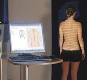
Rasterstereography: In clinical practice, this method is mainly used to study changes in the patient’s sagittal plane since it appears to be much more reliable in this plane than in the frontal plane. 

Moreover, current scientific evidence has failed to show the reliable correlation between diagnostic measurements between radiography (Cobb angle) and rasterstereography (1,2). We use rasterstereography to evaluate and monitor, over time, postural and structural problems affecting the sagittal plane, such as various forms of hyperkyphosis, long kyphosis, hyperlordosis, and so on.

This method is also very useful for evaluating the effectiveness of bracing or specific exercises over time.

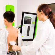
3D ultrasound imaging system: This is the first ultrasound imaging system capable of detecting and evaluating scoliosis. The only one currently available in Italy is at the Isico offices in Milan. 

Even though research (3) has shown very good correlations and agreements between ultrasound and radiographic measurements (respectively UCA – Ultrasound Curve Angle  and Cobb angle), the reliability of the system is not yet sufficient to allow the 3D ultrasound imaging system — and the same goes for rasterstereography — to replace radiography, which remains the diagnostic tool of reference for diagnosing scoliosis and for confirming its evolution.  

Based on our experience to date, we use the 3D ultrasound imaging system as a valuable ally in the frequent monitoring (every 3-4 months) of patients at increased risk of scoliosis progression, as it allows prompt detection of any worsening of the curves.

It should be recalled that this examination is suitable in patients with certain characteristics: e.g., for patients with a Risser sign of 0-1 who are undergoing either bracing or exercise-based treatment, for patients who are only being monitored for a possible scoliosis diagnosis, and finally for children over 5 years of age to reduce (annual) radiation doses/exposure.

Even though, according to current guidelines, standing anteroposterior and lateral-projection radiographs are the most reliable method for diagnosing scoliosis and sagittal deformities, both rasterstereography and 3D ultrasound imaging system can be considered valid and useful tools for monitoring the clinical condition over time, the first being used for more extensive assessments (of sagittal problems) and the second for targeted assessments (of scoliosis). Some authors suggest that they could be used for carrying out early screening in large populations (e.g., in schools) (2). 

1. Multicenter Comparison of 3D Spinal Measurements Using Surface Topography with Those From Conventional Radiography
DOI: 10.1016/j.jspd.2015.08.008

2. Is rasterstereography a valid noninvasive method for the screening of juvenile and adolescent idiopathic scoliosis?  DOI: 10.1007/s00586-018-05876-0

3. 3D ultrasound imaging provides reliable angle measurement with validity comparable to X-ray in patients with adolescent idiopathic scoliosis 10.1016/j.jot.2021.04.007

THE ROLE OF 3D ULTRASOUND IMAGING IN THE CLINICAL DIAGNOSIS AND MONITORING OF SCOLIOSIS DURING GROWTH

According to current guidelines, standing anteroposterior radiographs are the most reliable method for diagnosing scoliosis.
However, monitoring scoliosis, especially during growth, entails frequent radiation exposure (a full-spine X-ray every 6-12 months).
Even though there exists a low-dose X-ray system (EOS Imaging System), which we use with our patients, experts in recent years have been focusing on the quest to find an alternative, completely radiation-free system able to provide clinicians with equally reliable information.

Scolioscan®, developed in Hong Kong, is the first ultrasound imaging system that seems to offer valuable support in the clinical setting, both for monitoring scoliosis patients for progression of their curves and for screening populations for scoliosis.
Let’s take a closer look at it. Scolioscan® is a 3D ultrasound system that generates spine images using a volume projection imaging method. For this purpose, the patient’s back is scanned using a linear probe (10 cm wide, with an ultrasound frequency of 7.5 MHz) equipped with an electromagnetic spatial sensing device that allows three-dimensional spine reconstruction.  

The operator performs the scan freehand, using anatomical landmarks for reference: the scan starts from the sacrum (S1) and ends at the last cervical vertebra (C7).

It takes 30-60 seconds to perform, during which the patient has to be in a standing position, resting on four supports. At the end of the scan, the software immediately produces 9 images corresponding to progressively deeper coronal sections. The operator can thus select the image that gives the best view of the spinous and transverse processes of the vertebrae.
Thanks to these landmarks, the software reconstructs the curves based on the ultrasound spinous process angle (USSPA) or the ultrasound transverse process (i.e., lamina) angle (USLA) of each vertebra, identifying the slopes of the curves and the apical vertebrae. An ultrasound curve angle (UCA) is then calculated which measures the curves differently from how this is done on an X-ray. The system developers also worked out a mathematical formula for estimating the patient’s radiological Cobb angle. (1)
This examination is suitable for patients with a Risser sign of between 0 and 2, with one or two curves, and with a BMI of less than 23.
Research (2) has shown very good correlations and agreements between ultrasound (UCA) and radiographic (Cobb angle) measurements, with excellent intra- and inter-operator reliability.
However, more reliability is needed to allow ultrasound imaging to replace radiography, which remains the diagnostic tool of reference for diagnosing scoliosis and for confirming its evolution.  
The absence of radiation exposure is a massive advantage of this new system, allowing an accurate bone profile assessment. This, together with clinical measurements and evaluations, enables the specialist to decide on the course of the patient’s treatment, and can sometimes result in the decision not to perform a follow-up X-ray.  
This system can be used to monitor patients over time based on objective measurements and verify the effect of treatment in the short term, and it could — studies still need to confirm this as a means of helping patients optimise their self-correction movements.
At present, the application of this instrument is still in the exploratory phase, but this exploration should soon clarify all its clinical advantages. Based on our experience to date, Scolioscan® ensures that patients at greater risk of scoliosis progression get more frequent monitoring (even every 3 or 4 months), to help to detect any worsening.
Furthermore, Scolioscan® can also be considered a valid tool for preventive screening for scoliosis in school populations.

(1) Is radiation-free ultrasound accurate for quantitative assessment of spinal deformity in idiopathic scoliosis (IS): a detailed analysis with EOS radiography on 952 patients. Yi-Shun Wong, Kelly Ka-Lee Lai, Yong-Ping Zheng , Ultrasound in Med. & Biol., Vol. 00, No. 00, pp. 112, 2019

(2) 3D ultrasound imaging provides reliable angle measurement with validity comparable to X-ray in patients with adolescent idiopathic scoliosis. Timothy Tin-Yan Lee, Kelly Ka-Lee Lai, Jack Chun-Yiu Cheng, Journal of Orthopaedic Translation 29 (2021)