Posts

Do youngsters with scoliosis walk differently ?

In recent years, many studies have been conducted analysing gait and postural control during walking. Some of these have included gait analysis in youngsters affected by scoliosis.

The first thing to emerge from the literature is that the type of scoliosis does not influence gait. Indeed, no differences in the variables analysed were found between patients with adolescent idiopathic scoliosis and those with congenital scoliosis. Therefore, the gait disturbance in scoliosis is secondary to its existence and not related to its onset or aetiology [1].

In general, study results indicated that patients with scoliosis produced an asymmetrical rotation pattern in the frontal and transverse planes [2] and therefore, when walking, shifted their body to the side and rotated on themselves.

The position of the curve actually influences the motor pattern: in adolescent idiopathic scoliosis, patients with a single thoracic curve showed asymmetrical trunk movement in the transverse plane, whereas those with a single lumbar curve showed asymmetrical trunk movement in the coronal plane. These results suggest that the postural control strategy of patients with scoliosis differs according to the curve pattern [3].

Contrary to what X-rays deceptively show, scoliosis is not solely a deformity in the frontal plane but rather a fully three-dimensional deformity that X-rays can show only in two dimensions. This is confirmed by the findings of several analyses in which postural adjustments during walking were observed in all three space planes [4].

Scoliosis reduces the physiological curves in the sagittal plane, mainly due to loss of dorsal kyphosis. One study has, in fact shown that patients with scoliosis “may lean backwards and have posterior postural sway, which may be associated with hypokyphosis during walking” [5].

It has also been observed that adolescents with scoliosis have difficulty maintaining smooth bodyweight transfers during strides and that this difficulty increases with increasing severity of the spinal deformity. Gait parameters, namely the loading response, midstance phase and propulsion phase, were not found to differ between the left and right sides. However, patients with moderate and severe scoliosis had asymmetrical gait periods in both limbs,  suggesting abnormal asymmetrical gait patterns [6].

Given all this information, we were drawn to a recently published meta-analysis in which reduction of the stance phase and restriction of frontal pelvic motion was found to lead to prolonged activation timing of the quadratus lumborum, erector spinae and gluteus medius muscles [7].

A final question was whether these differences in walking might affect youngsters’ daily lives. In this regard, one study, using a pedometer installed on smartphones, showed no difference in daily walking distance between adolescents with and without scoliosis [8].

References

[1] Garg B, Gupta M, Mehta N, Malhotra R. Influence of Etiology and Onset of Deformity on Spatiotemporal, Kinematic, Kinetic, and Electromyography Gait Variables in Patients with Scoliosis-A Prospective, Comparative Study. Spine (Phila Pa 1976). 2021 Mar 15;46(6):374-382. doi: 10.1097/BRS.0000000000003796. PMID: 33620181.

[2] Yang JH, Suh SW, Sung PS, Park WH. Asymmetrical gait in adolescents with idiopathic scoliosis. Eur Spine J. 2013 Nov;22(11):2407-13. doi: 10.1007/s00586-013-2845-y. Epub 2013 Jun 4. PMID: 23732766; PMCID: PMC3886502.

[3] Nishida M, Nagura T, Fujita N, Hosogane N, Tsuji T, Nakamura M, Matsumoto M, Watanabe K. Position of the major curve influences asymmetrical trunk kinematics during gait in adolescent idiopathic scoliosis. Gait Posture. 2017 Jan;51:142-148. doi: 10.1016/j.gaitpost.2016.10.004. Epub 2016 Oct 11. PMID: 27764749.

[4] Wu KW, Wang TM, Hu CC, Hong SW, Lee PA, Lu TW. Postural adjustments in adolescent idiopathic thoracic scoliosis during walking. Gait Posture. 2019 Feb;68:423-429. doi: 10.1016/j.gaitpost.2018.12.024. Epub 2018 Dec 18. PMID: 30594870.

[5] Horng J, Liu XC, Thometz J, Tassone C, Duey-Holtz A. Evaluation of plantar pressures and center of pressure trajectories in Adolescent Idiopathic Scoliosis. Stud Health Technol Inform. 2021 Jun 28;280:131-135. doi: 10.3233/SHTI210451. PMID: 34190074.

[6] Zhu F, Hong Q, Guo X, Wang D, Chen J, Zhu Q, Zhang C, Chen W, Zhang M. A comparison of foot posture and walking performance in patients with mild, moderate, and severe adolescent idiopathic scoliosis. PLoS One. 2021 May 17;16(5):e0251592. doi: 10.1371/journal.pone.0251592. PMID: 33999943; PMCID: PMC8128255.

[7] Kim DS, Park SH, Goh TS, Son SM, Lee JS. A meta-analysis of gait in adolescent idiopathic scoliosis. J Clin Neurosci. 2020 Nov;81:196-200. doi: 10.1016/j.jocn.2020.09.035. Epub 2020 Oct 13. PMID: 33222916.

[8] Gaume M, Pietton R, Vialle R, Chaves C, Langlais T. Is daily walking distance affected in adolescent idiopathic scoliosis? An original prospective study using the pedometer on smartphones. Arch Pediatr. 2020 Aug;27(6):333-337. doi: 10.1016/j.arcped.2020.04.002. Epub 2020 Jun 17. PMID: 32563619.

Will the prominence disappear once the brace is on?

This question, together with others linked to aesthetic concerns, is often asked by parents and by youngsters about to start wearing a brace. After all, scoliosis is characterised by variable and sometimes marked clinical signs at the level of the trunk. The scoliotic prominence, linked to the spinal rotation component of the disease, becomes evident on bending forwards, but in some individuals it is also visible when standing. Changes in scoliosis have been found to be correlated with changes in the prominence, although this is not to say that a given degree of change will necessarily be proportional to a worsening or improvement of the hump; that said, a modification of the prominence can usually be taken as a sign that the scoliosis has changed.

Clinical monitoring of the prominence at medical checkups is therefore essential during brace treatment to establish whether there has been any reduction.

Bracing is a treatment that has two equally important objectives:

  • to stabilise or (when possible) improve the radiographic picture
  • to improve the appearance of the back

Essentially, the corrective force applied by the brace to the trunk reduces the rotation of the vertebrae, limits this particular contributing factor, and shapes the ribs and lumbar musculature to minimise the aesthetic impact of the curve. All these aspects will vary according to the type of brace, the experience of the orthopaedic technician who built it, the specific characteristics of the single spine and, last but certainly not least, the patient’s compliance with the treatment.

As we have said, clinical monitoring of the prominence at medical checkups is necessary, but the importance attached to this aspect should not be taken to mean that vertebral rotation always has an aesthetic impact and therefore constitutes an “enemy” to be fought at all costs.

In reality, in most cases, the problem is visible only to the trained eye of expert clinicians (doctors and physiotherapists), and parents and youngsters learn to see only it after the condition has been diagnosed and pointed out to them.

This is borne out by the fact that parents at the first consultation will often admit that, until then, they had never been aware of their child’s scoliosis and the associated aesthetic changes. For this reason, they usually have the impression that “the whole problem sprang up overnight”.

World Master: ready for the 2024 edition

First, some numbers confirm the idea of an international training event, which has now become part of Isico’s traditional educational offer, together with the Italian Master, the SEAS courses, and the course on the Sforzesco brace construction.
In 11 editions, there were 315 participants from 56 different countries, including 22 European countries, 17 Asian countries, 6 from the Americas, 4 from Africa, and 2 from Oceania. In other words, our World Master Course, which began in 2016, includes the whole world interested in the rehabilitation treatment of spinal pathologies.  

Participants have the privilege of attending the lessons of dozens of teachers among the leading international experts in rehabilitation treatment: “Improving my knowledge and skills about scoliosis, there was a lot of academic information presented in a systematic way. It made me read more, learn more, and learn about a different approach from other countries. It opened my mind to seeing scoliosis from a broader and more accurate perspective, which encourages me daily to learn more“. This is the comment of one of the participants in a past edition, Mohammed Firas Wahbeh, from Syria.  

Therefore, this is a learning opportunity that continues to be successful; in fact, we are close to opening registration for the IX Edition of the Online Master Course “Principles and Practice of Scoliosis Conservative Treatment – PPSCT”, which will start in January 2024.

How is the course organised?
The PPSCT course is delivered entirely online and self-administered through a dedicated online learning platform. It is divided into 16 modules, each lasting two or three weeks.
Every single module generally includes three recorded theoretical lectures (each about 45 minutes long), which participants can listen to at a time to suit themselves, enriched by scientific papers as in-depth material to complete the module task.
Discussion-group sessions complement the learning and the exchange with other students, focusing on specific aspects of the topic touched on within that specific module.

World Master: participants’ experiences
One of the most extraordinary aspects of these eight editions of our international online master course was to meet up and exchange views with students from all over the world who also told us how scoliosis is treated in their countries, what they expected from this training path, and what made the experience of this course unique.

We have mixed a short video of students’ feedback because no one better than those who have attended the course can tell others about it, together with a short presentation of the course given by Prof. Stefano Negrini, scientific director of our institute.

THE ROLE OF 3D ULTRASOUND IMAGING IN THE CLINICAL DIAGNOSIS AND MONITORING OF SCOLIOSIS DURING GROWTH

According to current guidelines, standing anteroposterior radiographs are the most reliable method for diagnosing scoliosis.
However, monitoring scoliosis, especially during growth, entails frequent radiation exposure (a full-spine X-ray every 6-12 months).
Even though there exists a low-dose X-ray system (EOS Imaging System), which we use with our patients, experts in recent years have been focusing on the quest to find an alternative, completely radiation-free system able to provide clinicians with equally reliable information.

Scolioscan®, developed in Hong Kong, is the first ultrasound imaging system that seems to offer valuable support in the clinical setting, both for monitoring scoliosis patients for progression of their curves and for screening populations for scoliosis.
Let’s take a closer look at it. Scolioscan® is a 3D ultrasound system that generates spine images using a volume projection imaging method. For this purpose, the patient’s back is scanned using a linear probe (10 cm wide, with an ultrasound frequency of 7.5 MHz) equipped with an electromagnetic spatial sensing device that allows three-dimensional spine reconstruction.  

The operator performs the scan freehand, using anatomical landmarks for reference: the scan starts from the sacrum (S1) and ends at the last cervical vertebra (C7).

It takes 30-60 seconds to perform, during which the patient has to be in a standing position, resting on four supports. At the end of the scan, the software immediately produces 9 images corresponding to progressively deeper coronal sections. The operator can thus select the image that gives the best view of the spinous and transverse processes of the vertebrae.
Thanks to these landmarks, the software reconstructs the curves based on the ultrasound spinous process angle (USSPA) or the ultrasound transverse process (i.e., lamina) angle (USLA) of each vertebra, identifying the slopes of the curves and the apical vertebrae. An ultrasound curve angle (UCA) is then calculated which measures the curves differently from how this is done on an X-ray. The system developers also worked out a mathematical formula for estimating the patient’s radiological Cobb angle. (1)
This examination is suitable for patients with a Risser sign of between 0 and 2, with one or two curves, and with a BMI of less than 23.
Research (2) has shown very good correlations and agreements between ultrasound (UCA) and radiographic (Cobb angle) measurements, with excellent intra- and inter-operator reliability.
However, more reliability is needed to allow ultrasound imaging to replace radiography, which remains the diagnostic tool of reference for diagnosing scoliosis and for confirming its evolution.  
The absence of radiation exposure is a massive advantage of this new system, allowing an accurate bone profile assessment. This, together with clinical measurements and evaluations, enables the specialist to decide on the course of the patient’s treatment, and can sometimes result in the decision not to perform a follow-up X-ray.  
This system can be used to monitor patients over time based on objective measurements and verify the effect of treatment in the short term, and it could — studies still need to confirm this as a means of helping patients optimise their self-correction movements.
At present, the application of this instrument is still in the exploratory phase, but this exploration should soon clarify all its clinical advantages. Based on our experience to date, Scolioscan® ensures that patients at greater risk of scoliosis progression get more frequent monitoring (even every 3 or 4 months), to help to detect any worsening.
Furthermore, Scolioscan® can also be considered a valid tool for preventive screening for scoliosis in school populations.

(1) Is radiation-free ultrasound accurate for quantitative assessment of spinal deformity in idiopathic scoliosis (IS): a detailed analysis with EOS radiography on 952 patients. Yi-Shun Wong, Kelly Ka-Lee Lai, Yong-Ping Zheng , Ultrasound in Med. & Biol., Vol. 00, No. 00, pp. 112, 2019

(2) 3D ultrasound imaging provides reliable angle measurement with validity comparable to X-ray in patients with adolescent idiopathic scoliosis. Timothy Tin-Yan Lee, Kelly Ka-Lee Lai, Jack Chun-Yiu Cheng, Journal of Orthopaedic Translation 29 (2021) 

Isico launches the second edition of the Sforzesco Course

After a first edition, organised in 2022, that saw participants from 13 countries worldwide, Isico is launching the second edition dedicated to the deepening and construction of its Sforzesco corset.

The combination of theoretical lessons presented by the Isico experts and the practical instructions given by the CPOs who collaborate with Isico are the winning points of this course, which recaptures the knowledge gained over 20 years of clinical practice with more than 10,000 patients wearing our Sforzesco braces.  
This second edition is entirely self-administered by the participants, and you can enrol at any time, without a deadline.
A live lesson will be held in December to summarise the material and address participants’ questions.  

Why joining? According to one of the participants in the last edition: “This training will be the starting point for the new chapter in our facility for the treatment of scoliosis. It will help me implement new strategies in current brace construction about the sagittal profile and in starting construction of the Sforzesco brace“. 
For more information and registration, visit https://en.isico.it/sforzesco-online-course/#top

Changing schools when you wear a brace

Every year, when the beginning of school comes round, we get asked the same question: “I’m changing school. I’m going to have new classmates and teachers. How should I tell them that I wear a brace?”

Well, the best thing is probably to start by telling the student who sits next to you, and then gradually tell the others. Don’t forget, it’s only a treatment, just like braces to treat teeth or glasses for eye problems.

It can be stressful trying to hide, and there’s absolutely no reason why you should! If your brace is well made it shouldn’t be noticeable under your clothes, although obviously if someone tries to tickle you or give you a hug, then they are going to feel your “shell”.  

Tell your friends about it: some of them might be a bit curious to begin with, others will want to help you with your backpack, and you will probably get teased a bit, but if you treat it as something quite normal then they will soon do the same!

It’s important to think carefully about what to wear for your first days at school. It’s best to choose clothes that make you feel good, and make sure you “wear” a big smile, too. Because even though a new school, like all new experiences, can be a bit scary, it will be exciting and interesting too!

What about those who tease you about your brace? Some classmates will tease you in a nice way, and if that’s the case, there’s no need to get offended. Of course, we all know that there can be some rather stupid people in this world, and wearing a brace is one way of working out who they are. Once you know, you will also know not to take any notice of them.

In fact, wearing a brace is a great way of learning not to care about how others may judge you. It will help you to make sure you only spend time with real friends, those who you know you can always count on! Unfortunately, nothing comes easy in life, and making friends takes time and effort, but you have to be prepared to persevere in order to see the results, just like with your brace!

The people who stick up for you and support you when things are tough are your real friends. As for the rest, take no notice of them. They are just people you happen to know!  

ISYQOL: the international version

With a pathology such as scoliosis that requires demanding treatment, at Isico we have known for years how fundamental it is to establish a relationship of trust with the patient and to have him adhere to the therapy, ensuring the possibility of a good quality of life in a brace. For this reason, years ago, a questionnaire was developed where our patients could express their assessment of the impact of wearing a brace on their daily lives.
From these questionnaires, the next step was to develop an online model made available to everyone, where, in ten questions, the patient himself evaluates his own well-being with regard to spinal pathologies (kyphosis, scoliosis, or other).
This is how the ISYQOL (Italian Spine Youth Quality of Life) questionnaire was born, which is based on the concerns expressed by patients and has proven to be particularly appropriate in patients with adolescent idiopathic scoliosis (AIS) who have not been treated surgically. 

In recent years, several studies have been published with adaptations and validations of the original Italian version in different languages. 
The latest revision is an international version published recently in the European Journal of Physical and Rehabilitation Medicine, a “Cross-cultural Validation of the Questionnaire: The ISYQOL International”.
The ISYQOL Italian version was translated into six languages using the forward-backwards procedure. The conceptual equivalence of the items’ content was verified, and any inconsistency was resolved by consensus. Finally, patients were asked to complete the ISYQOL before their consultation or treatment session without external help and without being influenced by their parents.
This work aims to assess the ability of ISYQOL to provide an accurate, cross-culturally equivalent measure of the quality of life for young people with idiopathic scoliosis from seven different cultures and languages (i.e. Canadian English, Canadian French, Greek, Italian, Polish, Spanish, and Turkish).
After this last revision, the questionnaire is now available in many languages. In addition to Italian and English, we have Chinese, Polish, Korean, Arabic, Persian, French, Spanish, and Greek versions.

A brace? Nothing to fear!

Unfortunately, scoliosis has to be treated in adolescence, which is already a very tricky and delicate phase in which youngsters often feel torn between wanting to be independent and wanting to fit in and belong.
We have all been through it and know how difficult this period of growing up can be. It’s not easy being the parent of an adolescent either. Sometimes we struggle to understand our children’s problems as they seem so far removed from our own. We might also struggle because we would like to be able to solve all their problems for them. This particular challenge, though, is one they need to overcome by themselves. What we, as adults, can do is be supportive, helping them to think things through and analyse situations, being careful never to minimise their problems or difficulties. 
For adolescents, it is very important to feel part of a group, and so anything that makes them feel “different” is scary. Years ago, youngsters worried about wearing braces on their teeth for this very reason, whereas nowadays so many have orthodontic treatment that no one bats an eye.   

Friendships are also important for teenagers, who will open up to one another, sharing their problems, feelings, insecurities and worries. True friends are the ones who support their brace-wearing peer (helping them to accept the device as a part of their daily life), and certainly not those who mock or tease another person for a health problem.

Also, as we all know, whenever we have what we consider to be a defect or flaw, we tend to see it as a far bigger problem than it really is. If, for example, we have a pimple on our face, however tiny, we become convinced that it’s the only thing everyone else notices about us!
In the same way, for some patients, a brace peeping out from under a t-shirt is an absolute disaster!
Compared with the braces of the past, today’s ones are made to be almost invisible under a vest or shirt. This is partly thanks to the orthopaedic technician’s skill and expertise and the specialist doctor who chooses the model. 

Over the years, we have seen that our patients can do seemingly impossible things with their brace on. Just take a look at the photos and videos published on our website, www.concorsetto.it, where you can find patients offering tips on how to conceal a brace under clothes, and others who do dance shows, go to the mountains or seaside, or do sport in their brace (even athletics, skiing and gymnastics).
Seeing other brace-wearing youngsters who have accepted the need to treat their condition and managed to deal with the situation in a positive way helps to normalise the whole experience!  Indeed, the key, basically, is to try and focus not on the inconvenience of a brace, but rather on the fact that spinal disorders have to be treated in order to prevent problems from arising in adulthood. It’s far better to pour your energies into fighting your scoliosis, rather than your brace. While you can’t change your brace, you can change your perception of it and how you approach it! In some cases, a patient might find it useful to talk to an expert, such as a psychologist, in order to overcome any difficulties in accepting the treatment.

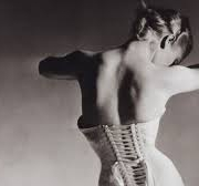
Recently, one young patient even sent us a photo showing the rock band Måneskin wearing corsets, which are not so different, in the end, from braces! Who knows, this could be the start of a new trend!
The last thing to remember is to take things step by step. Start by telling just a few close friends that you have started wearing a brace. That way, later on, with their help perhaps, you should find it easier to talk about it with others. Also, give yourself time to get used to it, as it is a whole new thing for you. In this way, slowly but surely, you will soon find you are getting on OK with your initially unwelcome new “friend”!

Can leg length discrepancy cause or worsen scoliosis?

Leg length discrepancy (LLD) is a common orthopaedic condition among children and adults, with a prevalence of 90% in the general population and 40% among athletes. LLD occurs when the lower limbs are of unequal length; the discrepancy is typically less than 10 mm and asymptomatic.
Individuals easily compensate for it through small adaptations of which they are very often unaware (such as bending or extending one leg more than the other). Some children are born with LLD; in other cases, the condition is acquired due
to causes such as tumours, radiation, infections or injuries.

LLD can be classified as functional or structural depending on the aetiology, i.e., the underlying cause.

Functional LLD refers to an apparent asymmetry, in the absence of real shortening or elongation of the osseous components of the lower limb. It is typically a result of pelvic obliquity related to adaptive soft-tissue shortening, joint or muscle contractures, ligamentous laxity, or axial misalignment. As the pelvis rotates, the legs are pulled into apparent different lengths.

Structural LLD, on the other hand, is a primary disorder causing actual physical shortening or lengthening of a limb, with the osseous change occurring between the ilium and the foot. The causes of shortening are more common than those of lengthening, and they may be congenital or acquired: shortening is most often due to fractures along the physis (Salter-Harris fractures), which can stop bone growth and lead to LLD.

LLD can be associated with several musculoskeletal disorders, including scoliosis and resultant degenerative spinal changes. However, the degree of LLD required to cause such disorders is still debated. LLD has been shown to cause pelvic obliquity in the frontal plane. In order to maintain shoulder balance and compensate for the pelvic obliquity, the condition that most frequently occurs is lumbar scoliosis with convexity directed towards the shorter limb. Scoliosis caused by LLD is not true scoliosis.

Should scoliosis due to LLD be treated?

This type of scoliosis, termed functional scoliosis, is non-progressive and involves a structurally normal spine with an apparent lateral curvature: it is evident in the erect position, but reduced when the subject is seated, supine or prone. The greater the degree of LLD, the more apparent the functional scoliosis may be. 

This type of scoliosis regresses fully or partially when its cause (i.e., the LLD) is removed. 

A simple remedy is to use shoe lifts, which may be internal or external depending on the degree of LLD.

If, after clinical evaluation, the functional scoliosis is found to be reducible (in part or totally) with a shoe lift, this solution will also be useful for reducing the overloading of the spinal joints and for reducing the structural and disc changes that occur over the years in the presence of scoliosis due to LLD (particularly when the difference between the two limbs is greater than 9 mm), as various studies have shown.

In a patient with structural and functional (LDD-related) scoliosis, it is appropriate to carry out tests to evaluate the usefulness or otherwise of a shoe lift. A shoe lift does not treat true scoliosis and the decision on whether or not to use it is a complex and individual one that should be made by the specialist.

Applebaum A, Nessim A, Cho W. Overview and spinal implications of leg length discrepancy: narrative review.  Clin Orthop Surg. 2021;13(2):127-34.

Giles LG, Taylor JR. Lumbar spine structural changes associated with leg length inequality. Spine (Phila Pa 1976). 1982;7(2):159-62.

Adams MA, Hutton WC. The effect of posture on the role of the apophyseal joints in resisting intervertebral compressive forces. J Bone Joint Surg Br. 1980;62(3):358-62. 

Murray KJ, Azari MF. Leg length discrepancy and osteoarthritis in the knee, hip and lumbar spine. J Can Chiropr Assoc. 2015;59(3):226-37.

Are bracing and exercises really still the only way to “solve” the problem?

This is a question many parents ask us when scoliosis is diagnosed. Perhaps their child has a particularly tricky curve, or maybe they themselves can remember dealing with the same condition when they were young. It is the same question we were recently asked by the mother of a five-year-old girl who, at this tender age, already has to reckon with a challenging treatment, “trapped” in a brace.

 Unfortunately, despite huge strides made in the formulation of less invasive and more effective braces (such as the Sforzesco type that we at ISICO, have long used as a valid substitute for plaster casts), bracing and specific exercises remain the only conservative treatments available for scoliosis.  

No parent wants their child to suffer the same negative experiences that they themselves remember, and it is perfectly understandable to be concerned about the possibility of them living the “nightmares” we did, and to want to protect them as far as possible. On the other hand, if this is something you have gone through yourself, you will actually be ideally placed to really understand all the difficulties your child is likely to face, and to help them find the best way to cope. It is not being complaining to ask whether other options exist and whether your child really does have to wear that uncomfortable piece of plastic — these are, after all, questions that any parent would ask.

 So, to return to the question, are there any other effective and less difficult treatments? Unfortunately, as explained in a recent study, manipulation and osteopathy, like all manual treatments, have not yet been shown to be effective (https://www.ncbi.nlm.nih.gov/pmc/articles/PMC9833903/).

Given the questions we so often get asked, we feel it is essential to properly explain the reason for certain aspects of the treatment, such as the need to ensure that brace-off hours are consecutive, the importance of performing specific exercises, and the duration and dosing of the treatment.

When deciding how long the treatment will last and how aggressive it needs to be in terms of the dose (i.e., number of brace-on hours per day), the factor we consider is the potential risk of the condition worsening. This risk is assessed based on the extent of the scoliotic curve and how much growing the patient still has to do (residual growth). Scoliosis worsens with growth, therefore the earlier it arises, the more likely it is to get worse, especially during growth spurts, when the youngster gains height rapidly (these usually happen at 6/7 years of age and in puberty).

Based on this information, the treatment can be adjusted, reducing or increasing the dosage according to the period of risk.

It also follows that the earlier scoliosis arises, the longer the treatment will need to be, given that bone growth has to be complete to reach a stable situation. Then there is the question of the “brace-on” hours. It has been demonstrated that after putting on a brace, it takes at least two hours for the spine to achieve the desired correction. 
This means that, when the brace is repeatedly taken off and put back on, we have to consider, and count, not only the brace-off hours, but also the hours needed for the spine to return to its correct position inside the brace. In the same way, every time the brace is removed the back tends to spring back to its starting condition (rather like a spring that has been pulled and then reverts to its original shape).

Then, the more frequently the brace is taken off to give the patient a break, the less effective the treatment will be compared with wearing it for the same number of hours but with fewer and longer breaks. For this reason, it is essential to try and establish and maintain a regular brace-wearing schedule.

We know that bracing treatment is very challenging and also that managing the brace-off hours can be difficult, especially during the summer. For this reason, we advise families, as far as possible, to choose somewhere cooler for their holidays, and also to talk to their specialist to see whether, on some days during the summer, the brace-on hours can be reduced or the brace-off time can be split into two blocks. Obviously, these decisions have to be taken on a case-by-case basis.

As for the specific exercises for scoliosis, these are designed to help support the back when the brace is not being worn, and also to prevent loss of muscle tone (otherwise an inevitable consequence of bracing). In the case of very young children (like the little girl whose mother prompted us to write this post), we recommend lots of sport to keep the muscles in shape, because children aged 4 to 6 years often don’t yet have the concentration necessary to be able to cope with exercise sessions.

Finally, in answer to the question, yes, at present, bracing and exercises are the only conservative therapy options that have been shown to work. This kind of treatment is certainly difficult and demanding, but we have to remember that it aims to help our children reach adulthood with healthy backs, and fortunately, we do at least have these “instruments” to offer them.