Different specialists, different prescriptions: how should we choose?

Most parents of a child with scoliosis embark on a similar journey: once they have received the initial diagnosis, they start consulting other specialists, seeking second or third opinions that might provide them with the confirmation and reassurance they need, and/or simply answers to further questions and doubts that have cropped up in the meantime. 

Often, though, parents who do this find themselves left with more questions than answers. This is because different specialists, faced with the same scoliosis case, can make different diagnoses and prescribe different courses of treatment.

Why is this? There may be different reasons. Before going any further, though, it is important to remember that only a specialist with specific training in vertebral pathologies can treat scoliosis. Once a timely and correct diagnosis has been made, it is necessary to decide how to treat the condition. 

The SOSORT guidelines on conservative treatment of scoliosis are an important resource in this regard. Taking into account the best current scientific evidence, as well as the extent of the curves and the degree of bone maturation, they provide suggestions on the most effective treatment. 

What the guidelines offer is not a single, specific course of treatment, but rather a series of options, ranging from the most conservative to the most aggressive, that could conceivably be prescribed in a given patient. 

However, science alone cannot meet all the needs of a long and complex course of treatment of the kind required in scoliosis. The evidence-based medicine approach brings together and combines scientific knowledge, the expertise and experience of the specialist, and the values and desires of the patient, and therefore makes it possible to formulate the most appropriate prescription for the individual case. 

Bearing all this in mind, then, it may well be that one doctor, considering the data collected during the examination and the discussion with the patient and the patient’s family, decides to prescribe a brace where another doctor might instead recommend only specific physiotherapy or even a wait-and-see approach, which consists of monitoring the situation for a few months to see how the scoliosis evolves.
These are very different prescriptions, but they are all valid. The patient will in any case be monitored following the prescription in order to make sure that the type of treatment, and the dose, are correct.
In this way, it is also possible to make any changes needed to avoid under-treatment (insufficient to contain the progression of the disease) or over-treatment (too taxing for the patient).

The big question remains: how do we parents go about choosing? There’s no easy answer. Given that our children will need to be on this therapeutic journey until they have finished growing, the important thing is to find someone we feel we can trust. In other words, we need to choose the specialist — and it must be someone with expertise in the conservative treatment of scoliosis — who we, and our child, felt to be the most reassuring and empathetic.

Once we have made our choice, we need to place our child’s care in the doctor’s hands. It is important to follow the instructions we are given, and not to change anything without the doctor’s agreement, as to do so could undermine the success of the treatment. 

Curves measuring less than 10 degrees: should we treat them?

As suggested by the Scoliosis Research Society (SRS), a scoliosis diagnosis is confirmed when a patient presents a Cobb angle measuring 10° or more and axial vertebral rotation. Maximum axial rotation is measured at the apical vertebra. (1) The SRS established this threshold in 1977, replacing the previous one of 7°. Ever since, 10 ° has conventionally been accepted, worldwide, as the threshold for diagnosing scoliosis.
However, structural scoliosis, with a potential for progression, can also be observed in the presence of Cobb angles measuring less than 10°. In fact, initial wedging of the vertebral bodies and disks can sometimes be registered with curves of 4°–7°. (2)

Idiopathic scoliosis, being a developmental disorder, most commonly arises and progresses during periods of accelerated growth (growth spurts).

The first such period occurs in infancy/early childhood, generally between 6 and 24 months of age, and the second between the ages of 5 and 8 years; finally, there is the pubertal growth spurt, which generally occurs at 11–14 years of age. (1)

Although the later stages of development are obviously not risk free, after puberty the rate of growth usually slows down, reducing the risk of progression of scoliosis. 

Can the risk of scoliosis progression be predicted in the case of curves measuring less than 10°?
There is, of course, always a chance that these curves will become more pronounced as the youngster grows, even, in some cases, to the point of requiring the use of a brace. But it is also true that most of them will remain stable over time without reaching the minimum criteria for a diagnosis of scoliosis. Certain factors may possibly be associated with an increased risk of scoliosis progression: a positive family history of scoliosis, laxity of ligaments, flattening of physiological thoracic kyphosis, a greater than 10° angle of trunk rotation (ATR), and growth spurts. All these factors should be evaluated by the attending physician. 

So, should we be treating these youngsters? In short, no. First of all, it is worth remembering, that the main aim of conservative treatment of scoliosis is to improve the patient’s appearance, but curves as mild as this rarely have an aesthetic impact; at most there may be some slight asymmetry of the trunk, but nothing that can be considered to exceed physiological parameters. With very rare exceptions, the only advice necessary in these cases is to opt for clinical monitoring of the patient, which can be considered to all intents and purposes a treatment, in the sense that it allows us to overcome the critical phases of development (which also correspond to the periods of greatest risk of progression of scoliosis) and also to intervene if any progression does occur. Monitoring is the first step in an active approach to idiopathic scoliosis, and it consists of clinical evaluations performed at regular intervals, ranging from every 2-3 months to every 36-60 months depending on the single case. 

In conclusion, any active treatment in this population of patients is actually overtreatment. Even just specific exercises, whose prescription constitutes first therapeutic step after monitoring alone, would cost these youngsters in time and effort, as well as being an economic cost.

A further aspect, not to be underestimated, is the psychological impact: starting a treatment amounts to confirming that the individual has a disease that needs to be treated, and this can lead them to start thinking of themselves as “sick”.

Furthermore, even though an exercise programme is not a particularly arduous undertaking, starting a treatment when there is no real need for one could compromise the youngster’s collaboration and commitment should a treatment be needed later on. This is an important consideration, because if their scoliosis does progress as they grow, specific exercises, rather than being useful, could become crucial, in order to avoid bracing for example.  

1 – 2016 SOSORT guidelines: orthopaedic and rehabilitation treatment of idiopathic scoliosis during growth

https://pubmed.ncbi.nlm.nih.gov/29435499/

2 – Radiographic Changes at the Coronal Plane in Early Scoliosis. Xiong, B., Sevastik, J. A., Hedlund, R., & Sevastik, B. (1994). Spine, 19(Supplement), 159–164. doi:10.1097/00007632-199401001-00008

https://pubmed.ncbi.nlm.nih.gov/8153824/

That hated hump!

In a person with scoliosis, forward bending of the trunk will cause a protuberance to appear on their back, at the level of the scoliotic curve: this anomaly is commonly referred to as a hump.

Scoliosis alters the alignment of the spine, and this alteration can be seen in the three dimensions of space: the affected vertebrae move sideways, taking on a different shape when viewed from the side; they also rotate, and this, especially if they are dorsal vertebrae, also causes a rotation of the rib cage. 

This hump is an aesthetic problem for people with scoliosis, as they fear that it is obvious to everyone else. But that is not the case at all. There is often a world of difference between the problem itself and how it appears in everyday life. 

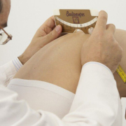
During a thorough postural analysis or a medical examination, the doctor or examiner will position the patient’s body to emphasise any asymmetries present precisely so that they can be identified and measured.

Nobody is perfectly symmetrical in daily life: when we move, we frequently twist our bodies and bend and rotate our joints, our movements involving different segments of our body in various combinations. As a result, we constantly develop asymmetries, humps, and combinations of bent and straight segments without even being aware of it.

Let’s take an example. A doctor examining a patient’s hump will have the youngster stand with his knees straight and body bent forwards so as to emphasise the protuberance on the side of the curve. Is it visible? Yes, if the patient’s scoliosis is sufficiently marked, it will be visible even to the untrained eye. 

Now, what if we ask someone with a healthy back to tie up their shoelace? To do this, most people will push their foot forward, bend their knees asymmetrically, and lean forwards, with their trunk deviating to one side (the side of the shoe needing to be tied). As they do this, their spine will be turned to one side, and a hump can be seen on their back, on the side of the leg, with the shoe needing to be tied. Does that hump mean they have scoliosis? Of course not! It is caused by them twisting their spine in order to reach their shoe.  

We can explain the scoliosis-induced asymmetry of the hips in a similar way. If we stand still with our feet parallel, our knees straight and our trunk aligned, our side and our hips will look symmetrical; in people with scoliosis, on the other hand, one hip will appear straighter in respect to the other. 

Now, let’s imagine how we normally stand. Do we ever actually stand with our feet positioned symmetrically and our bodyweight perfectly distributed between them? No! We usually stand with our weight on one foot and one hip thrust out. How do we look in this position? It is completely asymmetrical, but this asymmetrical appearance is due to our natural posture; it is not caused by scoliosis! 

Nobody ever notices these natural asymmetries because our bodies repeatedly assume them throughout the day. For this reason, even asymmetries caused by scoliosis are never really noticed by others. 

Going back to our “hump”: is it really such a bad thing, to the point that we should do everything in our power to get rid of it?

The back brace sometimes worn by youngsters with scoliosis, seeks to optimise the alignment of the vertebrae involved in the scoliosis curve, but how? It works by pushing “from the outside” directly on the affected portion to restore its symmetry. The force it exerts on the rib cage or on the soft tissues of the hips also affects the spine. 

 What is meant by an optimal alignment of the spine? The straightest possible? 

This isn’t an easy question to answer. Equally, it is not easy for the doctor to identify the correct balance of forces to be exerted on the spine by the brace pads. 

The best alignment will enable the treated spine to remain as stable as possible and to withstand the forces to which it is subjected in daily life. 

The strength of the spine depends to a large extent on its shape, as seen from the side. From this perspective, our spine is characterized by two curves, called lordosis and kyphosis. These curves must be well balanced: neither too pronounced nor too slight. 

Unfortunately, in scoliosis, and this applies particularly to dorsal scoliosis, the kyphotic curve is reduced, causing the back to appear flat or even hollow. In other words, the normal direction of spinal curvature is reversed, and the spine is weaker as a result. 

Unfortunately, the brace has no way of counteracting this problem, as all it can do is push. In fact, its corrective forces, which are applied to the hump to help align the vertebrae involved in the curve, have a flattening effect on the back’s upper part (dorsal section). 

Therefore, the doctor’s task is to find and maintain the best possible balance, considering the importance of all the planes of the spine.

What this means in practice is that it is sometimes necessary not to “go too far” in trying to eliminate the hump. Because if this objective can be achieved only by excessively reducing the dorsal kyphosis angle, the result will be a weaker and less healthy spine.  

Returning to physical activity after surgery for adolescent idiopathic scoliosis

Sport and physical activity are essential for psychosocial and physical well-being in children and adolescents, and youngsters with scoliosis are not subject to any limitations in this regard: they can do sports and other physical activities just like their peers can.

In severe scoliosis, i.e., in individuals with curves measuring more than 50° and presenting significant aesthetic and developmental deformities despite undergoing orthotic/bracing treatment properly administered according to the SOSORT criteria, surgery is indicated. 

“In some cases, patients who undergo corrective surgery for scoliosis may reduce their level of physical activity postoperatively due to a loss of spinal flexibility and pain. This inevitably impacts negatively on their quality of life – says dr Carmelo Pulici, physiatrist – No guidelines have yet been published on the resumption of physical activity after surgery for adolescent idiopathic scoliosis, and “expert opinion” is therefore all we have to rely on. Consequently, different surgeons may give different indications, depending on their experience and the particular type of approach”.

There are also individual factors at play, and the decision to resume physical activity may therefore depend on: the type of surgery performed, how the body responds and adapts to the metal rods inserted to correct the spine, the extent of the fusion, how recently the surgery was carried out, the characteristics of the patient, and also the type of sport practised (non-contact, contact or collision).  

For example, while some surgeons allow contact sports to be resumed six months after surgery, others recommend waiting a year, and some even rule out the resumption of collision sports altogether (https://pubmed.ncbi.nlm.nih.gov/26920125/). 

“In one study, return to sport/physical activities after surgery was examined using a specially created questionnaire (https://pubmed.ncbi.nlm.nih.gov/28604495/). By six months, most patients had returned to the activities they did before the surgery” – explains Dr Pulici. “The authors found that patients returned to athletics much earlier than expected. Despite this, there were no cases of loss of correction, implant failure or complications among the 95 patients included in the study”. 

A new surgical technique

In recent years, there has been a growing interest in vertebral body tethering (VBT), a new surgical technique used only in children and in early adolescence and, even then, only in carefully selected patients, given that the method is still experimental. VBT is proposed as a possible alternative to spinal fusion (currently the most widely used technique for the surgical treatment of scoliosis). Unlike spinal fusion, it does not reduce the mobility of the spine. 

“According to a recent study, VBT allows daily activities and sports to be resumed within three months of the surgery” Dr Pulici goes on. “Furthermore, most of the patients examined in the study reported an improvement in their athletic performance, and some were found to be doing more sports than before undergoing VBT”.

Even though surgeons today tend to allow patients to return to sports (even high-intensity ones) earlier than in the past (https://pubmed.ncbi.nlm.nih.gov/34267153/), we are still a long way from seeing a common approach to the management of postoperative resumption of physical activity in this patient population.

In the absence of guidelines, we at Isico recommend that patients turn to an experienced therapist in the post-operative period to ensure a gradual and safe return to physical activity, obviously following the instructions received from their surgeon.

It is important to avoid falling into the vicious cycle of a sedentary lifestyle. The less active you are, the less you feel inclined to do physical activity and the greater the risks to your health due to your lack of movement.

Scoliosis in the mirror: aesthetic concerns and psychological support

We are all critical when we look at ourselves in the mirror, and we tend to focus on and analyse the smallest detail, to the point that we sometimes get quite worked up about perceived flaws or defects that others probably don’t even notice.
This also applies to curves and asymmetries due to conditions like scoliosis. Indeed, to a young scoliosis patient these changes can appear extremely prominent, even when their friends and relatives insist that they can’t see anything wrong with the youngster’s back.

First of all, a brief preliminary remark. As part of scoliosis treatment, it is crucial to consider the appearance of the patient’s back.
This is not only because improving its overall aesthetics is one of the aims of the treatment (Negrini et al., 2018), but also because any change in its appearance, major or minor, can have a considerable psychological impact on the individual in question.

Of course, the way we see our own body and other people’s bodies is highly subjective, and aspects of our own body that bother us might not be the same aspects other people notice.

“For some scoliosis patients, the presence of asymmetries of the waist or shoulder blades, or a hump, will be an unbearable problem, whereas other patients may be quite unconcerned about them” says Dr Irene Ferrario, Isico psychologist. “And this can be entirely independent of objective clinical assessments; it may depend on the individual’s mood at the time, or on how much importance is attached to physical appearance in a given setting”. 

Sometimes, patients’ concerns about their appearance become disproportionate, turning into actual fixations that lead to them pouring far too much time and mental energy into working out how they can conceal their perceived defects.

“Having said that, minimising the patient’s concerns or trying to convince them that they don’t have any defects can, in some cases, be counterproductive” warns Ferrario. “Indeed, in the most severe cases, trying to appeal to reason to dispel concerns only has the effect of losing you the patient’s trust.”

Why seek psychological support

Therefore, it is advisable to seek help from a psychologist or psychotherapist in these cases. Unfortunately, many patients are reluctant to take this step as they remain convinced that removing the physical defect is the only way to resolve the problem and feel better. 

This is why scoliosis treatment must also address psychological needs to be effective.

On the one hand, specialists need to understand their patients’ distress and show them that they recognise the reasons for it. Once it is clear that a patient feels heard and understood, the therapist should then gradually and cautiously begin the process of formulating and presenting other possible reasons for the distress, helping the patient to know where it comes from and how it can be impacted by their environment and the people around them.   

“When we look at ourselves in a photo or in the mirror, we think we are seeing exactly what other people see when they look at us,” says Dr Ferrario, “but our eye is well trained to focus on the things we do not like about ourselves and thus to play on our insecurities. Whereas others see us as a whole and do not to dwell so much on specific details, we tend to analyse ourselves in minute detail, and our attention is always immediately drawn to the particular thing we are uncomfortable with.”

The main objective of psychological counselling, therefore, is to reduce, as much as possible, the discrepancy between the way we would like others to see us (our ideal self), how we think others should see us (self-guide), and how others see us (our real self).

“The course of treatment we do with our patients aims to help them rebuild their body image by encouraging greater awareness of their strengths and weaknesses” Dr Ferrario concludes. “Gradually, over time, patients manage to internalise this new image and eventually form a more realistic view of themselves. Patients also become more accepting of the aspects of their body that they used to see in a negative light”.

The Risser sign, growth and scoliosis: let’s clear a few things up

When patients come for medical consultations or physiotherapy sessions, numerous measurements get taken and recorded, often without less expert eyes even noticing.

On the other hand, other measurements are quickly seized upon, both by parents and youngsters. Take height, for example. The sliding piece barely has time to touch the patient’s head before the patient, hopeful, blurts out: “Have I grown? Can I leave off my brace now?”

Another milestone we are promptly informed of is menarche in girls, as parents are often convinced that when their daughters start their periods, they have finished growing, meaning that their treatment can come to an end. But this isn’t always the case. On the contrary, this delicate phase can sometimes coincide with the most marked progression of the disease, making it all the more important to act with caution.

Although these are two important examples of the many factors that need to be taken into account to work out what point a youngster’s growth has reached, it has been shown that increases in height and menarche do not necessarily coincide with the individual patient’s growth peak [1] and may therefore not be helpful and/or sufficient when it comes to deciding on the best course of treatment.

Since these manifestations are secondary growth characteristics, they can only be seen as an indication that the patient’s growth spurt has begun. What they do not tell us is precisely how far on it is. There is a scientific explanation for the traditionally held belief that girls “develop earlier” than boys. In fact, because testosterone starts to be released into the body after oestrogens, boys start their pubertal growth spurt later than girls.[1]

To manage scoliosis and optimise the treatment results of the condition, it is crucial to have a good idea of the patient’s residual growth potential and the time remaining until he/she reaches skeletal maturity. An accurate prediction of the growth rate is also required to know when the deformity is likely to be most at risk of progressing. On the other hand, once it has been established with certainty that the patient has finished growing, this is the time at which preventive measures can be stopped with only minimal risk of further deterioration of the curve. [1]

There are various methods we can use to evaluate bone growth in adolescence, and one of them is called the Risser sign.

An individual’s Risser grade can be determined from an anteroposterior X-ray of the spine. An advantage of this method is that the same X-ray can be used to measure both the number of Cobb degrees (necessary to diagnose scoliosis) and the degree of skeletal maturity, thereby limiting the patient’s radiation exposure.

From 0 to 5, Risser grades are assigned based on the amount of calcification present in the iliac apophysis, and the scale thus measures progressive ossification. A Risser grade 0 indicates a low degree of bone maturity: this status is present from birth through puberty.

A Risser grade 5 means that the iliac apophysis has fused to the iliac crest, and the structure is 100% ossified: this status is present in adults  [2]. 

It would be misleading to imagine the transition from Risser 0 to Risser 5 as a continuous and constant progression that occurs over a fixed time and at a set pace. This is because growth is not constant but proceeds at different rates in the different phases. There are times when it pauses, times when it speeds up considerably, and times when it slows down.

The crucial stage in a youngster’s growth, also vital for understanding the course of their scoliosis, is the pubertal growth spurt, during which the disease can alter the shape of the patient’s back in the space of just a few weeks. From the perspective of a Risser evaluation of skeletal maturity, this stage corresponds to the transition from Risser 0 to the complete acquisition of Risser 1.

Between Risser 2 skeletal maturity and the end of the Risser 3 stage, the growth spurt slows down, but as far as the scoliosis treatment is concerned, we still cannot lower our guard: the patient should continue to receive treatment.

Scoliosis treatment is brought to an end gradually as skeletal maturity increases. Once the patient has reached Risser grade 5 (complete skeletal maturity), the treatment can be terminated safely without fearing that some of the hard-won gains might be lost 

The Risser classification varies slightly in different parts of the world, with some differences found, in particular, between Europe and America. In Europe, the successive grades tend to be assigned more cautiously, in the sense that a patient is deemed to have passed from one stage to the next only in the presence of precise levels of bone maturation. On the other hand, the American tendency is to assign the successive grades sooner.

Another method for assessing skeletal maturity is the Sanders classification, whose eight grades are assigned based on the assessment of hand bone growth [3]. Some studies have found the Sanders classification more precise than the Risser sign. It shows higher staging sensitivity when growth is most rapid and is therefore more reliable during certain growth phases [4]. The problem with the Sanders classification is that it requires a separate X-ray of the hand, which therefore means that it could increase the patient’s radiation exposure.

All this information clearly shows that residual growth is essential to evaluate, but at the same time, difficult to establish and interpret.

Specialists can, of course, use the classification they prefer, which will be the one that, in their experience, works best for identifying and evaluating the growth peak in adolescence. It is essential that they can correctly interpret all the data they collect, including from radiographs and patients themselves, to optimise the timing and results of the treatment. 

References

[1] Cheung JPY, Luk KD. Managing the Pediatric Spine: Growth Assessment. Asian Spine J. 2017 Oct;11(5):804-816. doi: 10.4184/asj.2017.11.5.804. Epub 2017 Oct 11. PMID: 29093792; PMCID: PMC5662865.

[2] Greiner KA. Adolescent idiopathic scoliosis: radiologic decision-making. Am Fam Physician. 2002 May 1;65(9):1817-22. PMID: 12018804.

[3] Sanders JO, Khoury JG, Kishan S, Browne RH, Mooney JF 3rd, Arnold KD, McConnell SJ, Bauman JA, Finegold DN. Predicting scoliosis progression from skeletal maturity: a simplified classification during adolescence. J Bone Joint Surg Am. 2008 Mar;90(3):540-53. doi: 10.2106/JBJS.G.00004. PMID: 18310704.

[4] Minkara A, Bainton N, Tanaka M, Kung J, DeAllie C, Khaleel A, Matsumoto H, Vitale M, Roye B. High Risk of Mismatch Between Sanders and Risser Staging in Adolescent Idiopathic Scoliosis: Are We Guiding Treatment Using the Wrong Classification? J Pediatr Orthop. 2020 Feb;40(2):60-64. doi: 10.1097/BPO.0000000000001135. PMID: 31923164.

Adults: can hyperkyphosis be improved?

With the passing years, many adults start to realise, when they look at themselves in the mirror, that they are getting increasingly stooped. Some people are unwilling to accept this situation and start wondering whether they can do anything to arrest this process. The question is, can this condition be improved or is it pointless even to try?

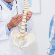
The condition we are talking about is HYPERKYPHOSIS. If you look at a person sideways on, you see that their back is not straight, but has natural curves, whose function is to cushion the forces that act on the spine. Following the back line from the top down, we see that first, at cervical level, there is a forward curvature, termed LORDOSIS, then a backward dorsal one, called KYPHOSIS, followed by another forward curve, at lumbar level, also called LORDOSIS. When the amplitude of the dorsal kyphotic curve, measured on an X-ray, exceeds the normal range, we speak of HYPERKYPHOSIS. Usually, this curve measures between 20 and 60 Cobb degrees.
Various factors explain this considerable range. Some are positional and related to the type of examination performed (for example the position of the arms), while others are linked to the associated disorder itself, which may be characterised by marked (e.g., scoliosis) or more prominent (e.g., idiopathic hyperkyphosis, Scheuermann’s disease) curves. Elderly people often present hyperkyphosis caused by the osteoporotic vertebral collapse. As the bones become more fragile, even minor movements can cause tiny fractures of the anterior portion of the vertebrae, resulting in progressive bending of the whole back

Everyone’s back bends forward more as the years go by, regardless of whether or not they have hyperkyphosis.
Why is this?  Most people spend much of their time, i.e., many hours of most days over many years, in a hunched position, with the head looking downwards. In fact, in our daily lives, we are often in the sitting position, which encourages forward flexion of the back; furthermore, many of the activities that require us to move around (cooking, cleaning, DIY, hobbies) also involve bending forwards. For all these reasons, dorsal kyphotic curves tend to get progressively worse over time, we become increasingly stiff, and the trunk extensor muscles grow weaker, resulting in postural collapse. In short, all these factors, combined, leave us “crushed” by the force of gravity.

What are the effects of hyperkyphosis? In adults and the elderly, hyperkyphosis can increase our risk of back pain and worsen our quality of life as we find it increasingly challenging to support our back, both when seated and when standing. Another effect is impaired balance and stability when walking.

So, to go back to our original question: is it possible to break this vicious cycle through physiotherapy and, in particular, through specific exercises? 

The answer is yes! The initial objectives of the treatment are to reduce the stiffness of the dorsal spine and strengthen the trunk muscles that oppose the force of gravity, so as to facilitate postural recovery, and to integrate the correction into daily life. Indeed, from the outset, the treatment approach based on specific exercises encourages patients to learn the crucial “self-correction” movement that allows them to achieve optimal realignment of the spine in the sagittal plane without compensating for this at other levels of the spine (reference: Exercise for improving age-related hyperkyphosis: a systematic review and meta-analysis with GRADE assessment. Ponzano M, Tibert N, Bansal S, Katzman W, Giangregorio L. Arch Osteoporos. 2021 Sep 21;16(1):140. doi: 10.1007/s11657-021-00998-3. PMID: 34546447)

.

Once the improvement has been obtained, it needs to be made stable and lasting. This involves reducing the frequency of the specific exercises and integrating them with other types of physical or sporting activity, all the time continuing to maintain the correction in daily life.  

Together, all this translates into less pain, better physical function and a more attractive back.

Can bracing improve trunk asymmetries in young adults?

Scoliosis is a condition characterised by the presence of a vertebral deformity in the three planes of space. The extent to which this deformity is visible externally varies from case to case. As stated in the guidelines recently developed by SOSORT, one of the primary objectives of scoliosis rehabilitation treatment is to improve trunk asymmetries. Achieving this objective has important implications for quality of life — after all, having a positive self-image helps to ensure a good level of psychological wellbeing and this, in turn, translates into a better quality of life.

Not uncommonly, adolescents with scoliosis lack the willpower or motivation to undertake bracing treatment, or perhaps have never had a medical specialist recommend it to them; others, quite simply, may not even have realised that they had scoliosis.
Basically, many different factors can affect what scoliosis treatment a person received, or did not receive, in adolescence. But the fact is that some people reach adulthood before realising, for example, that they have one waist straighter than the other, that part of their rib cage protrudes, or that one breast is higher than the other.

What can be done to improve these asymmetries?

“Although there is no scientific evidence available to help us answer this question with precision, we at ISICO have performed some bracing treatments in young adults (up to 25 years of age)explains dr Fabio Zaina, physiatrist – In such cases, our doctors, during the consultation, are clear from the outset: this is a long and demanding course of treatment. For the first few months, the brace should be worn full time, i.e., for 23 hours a day, after which the brace-wearing time is reduced very gradually in an effort to maintain the results obtained”.

Patients wanting to start bracing must be absolutely convinced about it and highly motivated, too, because any sudden interruption of the treatment, without respecting the abovementioned weaning-off phase, can negatively affect the stability of the spine.

On the subject of stability, we should also point out that bracing treatment must always be combined with specific exercises based on self-correction. The purpose of these exercises is to keep the back muscles strong and teach the patient how to keep their trunk correctly positioned during the various activities of daily life.

Can adults obtain a radiographic improvement?

Unfortunately, radiographic improvements cannot be achieved in adulthood. In individuals who have reached full skeletal maturity, bracing is never proposed with this aim in mind.

In other words, bracing in adults can improve the external asymmetries related to the scoliotic curves, but it cannot reduce the Cobb degrees of the curves themselves.

Are there no other options available for adults who are reluctant to wear a brace?  

“Adults can always follow a specific physiotherapy programme based on self-correction exercises explains Valentina Premoli, physiotherapist- These exercises are a way to act on the postural component of scoliosis. They serve to counteract the spine’s tendency to collapse to one side, and can thus improve the functioning of the back generally. This type of treatment helps to limit the worsening of the asymmetries that accompanies the spine’s tendency to give way in the direction of the curves — a tendency that becomes increasingly marked as we get older”.

Scoliosis and pregnancy

For a woman, discovering she is pregnant is often one of the most memorable, most exciting and happiest moments in her life. Thinking about the baby, imagining it and talking about it, not to mention feeling it inside her, arouses a number of precious and positive emotions: hope, tenderness and love. However, at the same time, pregnancy leads to various changes, in her body, her self-image and her vision of the future.
Furthermore, she will need to make adjustments and seek new balances in her (often busy) daily life, her rhythms and her relations with others. 

All this can generate normal and entirely understandable fears, and these can be amplified in mothers-to-be who happen to be affected by a condition like scoliosis. Many such women will already have expressed anxiety over their ability to conceive, carry and give birth to a child.
Pregnancy and childbirth, on account of the physical demands they make, can indeed be quite a daunting prospect for these women.

An interesting recent review of the literature (Dewan MC, Mummareddy N, Bonfield C. The influence of pregnancy on women with adolescent idiopathic scoliosis. Eur Spine J. 2018 Feb;27(2):253-263. doi: 10.1007/s00586-017-5203-7. Epub 2017 Jun 29. PMID: 28664223.), focusing on the interaction between pregnancy and scoliosis, examines these very issues. Just to give an idea, in numerical terms, of the analysis carried out, this review included 134 articles and examined 22 studies, referring to a total of 3125 patients.

First of all, the review considered whether and how scoliosis affects the timing and outcomes of pregnancy. It would appear that women with idiopathic scoliosis need not worry about their possibility of having children, even though they have a slightly lower probability of becoming pregnant compared with age-matched women, and may be slightly more likely to receive fertility treatment. Furthermore, women with scoliosis, regardless of whether they underwent surgery or bracing treatment, can expect to have a similar number of children as healthy women. 

However, the studies considered have certain limitations: most of them failed to specify whether the women with scoliosis had been actively seeking or desired pregnancy. Similarly, it is not clear whether all the patients were followed up until menopause. Furthermore, marriage rates, often not even mentioned, were not uniform across the studies.
In the absence of indications on these aspects, the slightly higher rate among women with scoliosis who do not have children could be misinterpreted.

Bracing + exercises + adherence to treatment = a recipe for successful treatment!

Idiopathic scoliosis is a disease that causes abnormal growth of the spine. Once a spinal curve has appeared, the vertebrae of a young patient are liable to become deformed in the three planes of space. This risk persists until he or she has finished growing.
Through conservative treatment, we aim to ensure that our patients, as adults, will have a strong back. In other words, we want them to reach adulthood with a healthy and functional spine, and that does not necessarily mean a perfectly straight one!

Therapy is therefore undertaken in order to try and limit the natural worsening of scoliosis curves through treatments proportionate to the severity of the condition.

To this end, there exist two main methods: specific physical exercises and bracing, and both need to be applied correctly and with the necessary adherence to treatment.

To decide whether a young patient needs to be prescribed a brace, the treating physician considers a series of factors, for example the size of the curve, the patient’s bone age, and whether or not he/she presents asymmetries (of hips, pelvis, scapulae, shoulders and so on).

When bracing is deemed warranted, we urge our youngsters to adhere scrupulously to the instructions given in order not to compromise the effectiveness of the treatment, and indeed to obtain the best possible correction.
This means that they must fasten and tighten their brace correctly, as shown by our doctors during the testing phase. If they do this, their brace will be less visible under their clothing; it will also be less prone to move about when they are walking, running and even sitting, and therefore more comfortable.

In addition to being more visible under clothes, a brace that is worn too loosely is less effective: it will not give the results that would have been achieved by wearing it properly.

According to data we have gathered, bracing treatment should produce its most marked results in the first months, and adhering to the prescribed number of brace-on hours is what makes the difference in this regard.

Bracing treatment is always prescribed together with specific physical exercises, i.e., 15-20 minutes per day of spinal self-correction and stabilisation exercises that help patients get into the habit of regularly correcting their posture themselves, even during the hours they are permitted to leave their brace off. 

We constantly remind our patients that whenever they remove their brace, self-correction becomes all important, and that “voluntary and active” self-correction works just like the brace itself does. Clearly, though, this demands good self-awareness on their part and a willingness to collaborate.

In “Specific exercises performed in the period of brace weaning can avoid loss of correction in Adolescent Idiopathic Scoliosis (AIS) patients: Winner of SOSORT’s 2008 Award for Best Clinical Paper”, we  showed that patients who regularly did specific exercises obtained a stable result when they finally stopped wearing their brace. Instead, those who did specific exercises discontinuously showed a worsening of a few degrees, while the curves of those who did no exercises at all worsened by an average of 10 Cobb degrees or more.

And it is important to remember that all these patients were prescribed the same type of brace, and the same number of bracing hours.

Brace weaning, which is carefully monitored by the physician, must be done extremely gradually, as the spine needs time to adapt to the absence of an external support. This is particularly true in the case of patients treated for very severe curves. During this delicate phase, self-correction exercises become even more important, helping to prevent the spine from once again collapsing in the direction of the curve. 

We often say that strength comes from within: a patient’s own determination to wear his/her brace consistently and do his/her exercises correctly will together help to ensure that conservative treatment is a success – and it is important to understand correction of the scoliotic curve is not the only measure of success. Success also means stabilising the curve and curbing its tendency to worsen as the patient grows.