Rastereography vs 3D ultrasound imaging system: when should we choose one instead of the other?
Let’s start with the thing they have in common: to lower radiation exposure. Indeed, these two methods, although unable to accurately reproduce the Cobb angle, were both created to reduce the radiation exposure of patients being monitored for spinal disorders, especially during pubertal growth.
Non-invasiveness and absence of radiation exposure are huge advantages of these methods, used for evaluating the curves of the spine, physiological and otherwise; combined with clinical measurements and evaluations, they allow the specialist to decide on the course of the patient’s treatment. And in many cases without the need for a follow-up X-ray.
We have already explored the features and peculiarities of these methods in one previous post and another one
So, when should we opt for one as opposed to the other?
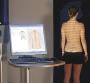
Rasterstereography: In clinical practice, this method is mainly used to study changes in the patient’s sagittal plane since it appears to be much more reliable in this plane than in the frontal plane.
Moreover, current scientific evidence has failed to show the reliable correlation between diagnostic measurements between radiography (Cobb angle) and rasterstereography (1,2). We use rasterstereography to evaluate and monitor, over time, postural and structural problems affecting the sagittal plane, such as various forms of hyperkyphosis, long kyphosis, hyperlordosis, and so on.
This method is also very useful for evaluating the effectiveness of bracing or specific exercises over time.
3D ultrasound imaging system: This is the first ultrasound imaging system capable of detecting and evaluating scoliosis. The only one currently available in Italy is at the Isico offices in Milan.
Even though research (3) has shown very good correlations and agreements between ultrasound and radiographic measurements (respectively UCA – Ultrasound Curve Angle and Cobb angle), the reliability of the system is not yet sufficient to allow the 3D ultrasound imaging system — and the same goes for rasterstereography — to replace radiography, which remains the diagnostic tool of reference for diagnosing scoliosis and for confirming its evolution.
Based on our experience to date, we use the 3D ultrasound imaging system as a valuable ally in the frequent monitoring (every 3-4 months) of patients at increased risk of scoliosis progression, as it allows prompt detection of any worsening of the curves.
It should be recalled that this examination is suitable in patients with certain characteristics: e.g., for patients with a Risser sign of 0-1 who are undergoing either bracing or exercise-based treatment, for patients who are only being monitored for a possible scoliosis diagnosis, and finally for children over 5 years of age to reduce (annual) radiation doses/exposure.
Even though, according to current guidelines, standing anteroposterior and lateral-projection radiographs are the most reliable method for diagnosing scoliosis and sagittal deformities, both rasterstereography and 3D ultrasound imaging system can be considered valid and useful tools for monitoring the clinical condition over time, the first being used for more extensive assessments (of sagittal problems) and the second for targeted assessments (of scoliosis). Some authors suggest that they could be used for carrying out early screening in large populations (e.g., in schools) (2).
1. Multicenter Comparison of 3D Spinal Measurements Using Surface Topography with Those From Conventional Radiography
DOI: 10.1016/j.jspd.2015.08.008
2. Is rasterstereography a valid noninvasive method for the screening of juvenile and adolescent idiopathic scoliosis? DOI: 10.1007/s00586-018-05876-0
3. 3D ultrasound imaging provides reliable angle measurement with validity comparable to X-ray in patients with adolescent idiopathic scoliosis 10.1016/j.jot.2021.04.007

