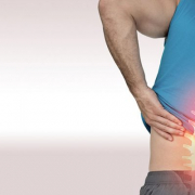
Posts

Disc herniation in youngsters : what it is and how to prevent it

Three out of ten people suffer from persistent troublesome pain in the lower back that sometimes radiates even as far as the lower limbs, a pain that lasts for a year or more rather than disappearing in two or three weeks as is normally the case. Commonly referred to as “sciatica” or “lumbosciatica”, it is due to a condition called lumber disc herniation, which is a common cause of pain, including low back pain, and can significantly impact the quality of life of those affected. Unfortunately, this problem is now also seen in youngsters, too: “Over the years, changes in kids’ lifestyles — they spend increasingly more time sitting down and less and less in movement — have gone hand in hand with a growing prevalence of certain diseases, including lumbar disc herniation,” explains physiotherapist Sara Rossi Raccagni.

Let’s examine more closely what we are talking about. When material from the nucleus pulposus of an intervertebral disc is pushed through a tear in the fibrous ring (annulus) that forms its wall, and therefore out of its natural location, the disc is described as herniated. The intervertebral discs are the cushions located between the vertebrae, which absorb the loads to which the spine is subjected in the course of the day. The discs most affected by this degenerative process are the ones located between the fourth and fifth lumbar vertebrae and between the fifth lumbar vertebrae and the sacrum.

What are the risk factors that may contribute to the occurrence of disc herniation?

A recent study (Risk factors for lumbar disc herniation in adolescents and young adults: A case-control study ) investigated risk factors for lumbar disc herniation in 208 patients aged up to 25 years.

Obesity, which corresponds to a BMI (body mass index) greater than 30, has been found to be closely associated with the risk of lumbar disc herniation, probably due to the greater load exerted on the structures of the spine.

Poor posture when seated and spending more than six hours a day seated have both been identified as risk factors for the onset of disc herniation: “So-called lazy postures (sitting slouched back in a chair with your pelvis slid forward, or sitting bent over with your head and upper body leaning forward) put the spine under extra stress and increase the likelihood of disc degeneration, especially when they are maintained for a long time” Rossi Raccagni goes on. “Prior low back traumas also seem to increase the risk of disc herniation, especially in athletes because of the greater stress exerted on the structures of the spine. This applies especially when training (and the initial warm-up in particular) is not done correctly”.

Finally, another risk factor to bear in mind is heredity, in other words, whether there are cases of herniated disc in the family that could be linked to genetic anomalies, i.e., mutations in genes coding for extracellular matrix components, inflammatory markers and protein metabolism.

With the exception of the latter risk factor, on which we cannot intervene, all the others can be controlled by adopting a healthy lifestyle, reducing as much as possible the time spent sitting down and doing regular exercise as appropriate for your physique.

To reduce the risk of accidental trauma, it is a good idea to warm up completely before starting any sport-specific training. This warm-up increases the elasticity of your muscles and your spinal range of motion in all directions. 

If you have a herniated disc, it is always advisable to consult doctors who specialise in the treatment of spinal disorders, as they can guide you step by step and also put you in the safe hands of expert physiotherapists. 

The first-choice treatment is the type termed “conservative”. Conservative treatment must always be multidisciplinary and can include the use of drugs (anti-inflammatory and neurotrophic drugs), postural education, cognitive-behavioural therapy, manual therapy and exercises” Rossi Raccagni concludes. “If the pain fails to improve with conservative therapy or neurological deficits arise (stenosis and/or marked sensory deficits), then the patient might be referred for surgery.

Disc herniation can also be treated using “minimally invasive” methods, which are considered to fall mid-way between conservative and surgical treatments. They include oxygen-ozone therapy and epidural injections.

“I have had scoliosis surgery. Do I face any risks with pregnancy?”

Let’s imagine the case of a healthy, active young woman who, as a teenager, had challenging scoliosis treatment, to the point of requiring surgery. Now, a few years on, she faces a new challenge: she wishes to become pregnant in order to fulfil her dreams of becoming a mother. 

Pregnancy is a period that naturally brings worries as well as lots of new information that needs to absorbed in order to be able to enjoy this special time. Those with scoliosis might start wondering “Will I have problems in pregnancy because of my scoliosis and the surgery I had? Or the reverse: “Could pregnancy aggravate my back condition?”.

Let’s try and help this mother-to-be, by providing answers based on the best available scientific evidence.

Several studies in the literature have investigated the topic “Pregnancy and surgery”, examining how one might influence the other.
Let us begin by underlining one reassuring aspect: as far as has been demonstrated so far, pregnancy (be it one pregnancy or more) has no consequences (in terms of a progression or deterioration of the curve) either on the surgically fused portion of the spine or on the vertebrae that were left free (https://pubmed.ncbi.nlm.nih.gov/32272267/https://pubmed.ncbi.nlm.nih.gov/2948962/). 

Nevertheless, it is always a good idea, when possible, to seek the opinion of the surgeon who performed the surgery, in order to have answers to your queries and precise instructions to follow.

Moreover, if the patient chooses epidural pain relief and/or chooses or requires to deliver by caesarean section with epidural anaesthesia, the anaesthetist might wish to avoid the surgically-treated portion of the spine (https://pubmed.ncbi.nlm.nih.gov/32578160/ ; https://pubmed.ncbi.nlm.nih.gov/30610987/ ), even though this decision by the medical team, made in agreement with the patient, seems to be taken more as a precaution.

In general, the use of epidurals in women with previous scoliosis surgery is comparable to what is observed in scoliosis patients who have not had operative treatment. Furthermore, according to the available data, anaesthesiologists seem reluctant to perform an epidural if the surgically treated portion of the column is below the third lumbar vertebra, preferring instead to opt for general anaesthesia (https://pubmed.ncbi.nlm.nih.gov/26131384/ ; https://pubmed.ncbi.nlm.nih.gov/32578160/https://pubmed.ncbi.nlm.nih.gov/30610987/ ).

With regard to possible pain or complications during pregnancy and delivery, some studies have shown that there are no differences between women who have and those who have not had scoliosis surgery (https://pubmed.ncbi.nlm.nih.gov/9391799/ ).
On the other hand, low back pain during pregnancy seems to be more frequent in surgically treated patients, although it disappears relatively quickly after delivery. ( https://pubmed.ncbi.nlm.nih.gov/30610987/ ).
All in all, then, a mother-to-be can face this exciting new chapter in her life with complete peace of mind, even though she should not forget to seek the opinion, regarding her back, of both her family doctor and a spine specialist. In this way, it is possible to prevent any pain and have the best possible experience of pregnancy and childbirth

Low back pain

Every year, the Italian Scoliosis Study Group selects the best published papers on conservative spine treatment from the global scientific literature.
Here is the abstract from one of these papers. 

Long-term follow-up of untreated Scheuermann’s kyphosis
  Enrique Garrido, Simon B Roberts, Andrew Duckworth, Joseph Fournier 
PMID: 34212306 DOI: 10.1007/s43390-021-00354-y

Abstract

Study design: Long-term cross-sectional study.

Objectives: To investigate the long-term effects of untreated Scheuermann’s kyphosis on quality of life, and its relationship to radiographic parameters of spinal deformity. Previous studies reported reduced self-image, increased pain and impaired physical status. Little is known of the long-term impact of sagittal plane deformity in untreated SK.

Methods: One hundred and thirteen consecutive untreated patients with SK were identified from a national service database prior to 2000, when surgery was not offered at this unit. 81 of these patients were available for evaluation; 66 (81%) consented to questionnaire and clinical evaluation, and 47 (58%) consented to additional radiological evaluation. Health-related quality of life (HRQoL) was compared to normative population values. Mean age was 45.1 years (31-65), and mean follow-up was 27 years (16-36). 57 patients had thoracic kyphosis and 9 had thoracolumbar deformity.

Results: SRS-22 and SF-36 scores were lower, and ODI was greater in patients with untreated SK compared to normative population values. Kyphosis progressed from mean 66° at skeletal maturity to 78° (p < 0.001) after mean follow-up of 27 years. Long-term progression of untreated SK was 0.45°/year (n = 47). Multilinear regression showed good correlation between increasing SVA and worse ODI scores (r = 0.59; p = 0.001). Increasing SVA also correlated with worse function, pain and mental health scores reported by SRS-22, and with worse physical function and bodily pain scores reported by SF-36. Increasing CL correlated with worse SF-36 physical function scores. Increasing cSVA and increasing TK correlated with worse SRS-22 self-image scores.

Conclusion: SRS-22 and SF-36 scores were lower, and ODI was greater in patients with untreated SK compared to normative data. Long-term progression of untreated SK was 0.45°/year (n = 47). Increasing SVA correlated with worse SF-36 physical function, SRS-22 function, SRS-22 pain and higher ODI scores. Total kyphosis (TK) and cSVA were independent predictors of low SRS self-image.

Level of evidence: III.

Keywords: Disease; Kyphosis; Natural history; Outcome; Scheuermann’s.

https://pubmed.ncbi.nlm.nih.gov/34212306/

Low back pain

Every year, the Italian Scoliosis Study Group selects the best published papers on conservative spine treatment from the global scientific literature.
Here is the abstract from one of these papers. 

Low back pain
 Nebojsa Nick Knezevic, Kenneth D Candido, Johan W S Vlaeyen, Jan Van Zundert, Steven P Cohen
PMID: 34115979 DOI: 10.1016/S0140-6736(21)00733-9

Abstract

Low back pain covers a spectrum of different types of pain (eg, nociceptive, neuropathic and nociplastic, or non-specific) that frequently overlap. The elements comprising the lumbar spine (eg, soft tissue, vertebrae, zygapophyseal and sacroiliac joints, intervertebral discs, and neurovascular structures) are prone to different stressors, and each of these, alone or in combination, can contribute to low back pain. Due to numerous factors related to low back pain, and the low specificity of imaging and diagnostic injections, diagnostic methods for this condition continue to be a subject of controversy.
The biopsychosocial model posits low back pain to be a dynamic interaction between social, psychological, and biological factors that can both predispose to and result from injury, and should be considered when devising interdisciplinary treatment plans.
Prevention of low back pain is recognised as a pivotal challenge in high-risk populations to help tackle high health-care costs related to therapy and rehabilitation. To a large extent, therapy depends on pain classification, and usually starts with self-care and pharmacotherapy in combination with non-pharmacological methods, such as physical therapies and psychological treatments in appropriate patients.
For refractory low back pain, a wide range of non-surgical (eg, epidural steroid injections and spinal cord stimulation for neuropathic pain, and radiofrequency ablation and intra-articular steroid injections for mechanical pain) and surgical (eg, decompression for neuropathic pain, disc replacement, and fusion for mechanical causes) treatment options are available in carefully selected patients.
Most treatment options address only single, solitary causes and given the complex nature of low back pain, a multimodal interdisciplinary approach is necessary. Although globally recognised as an important health and socioeconomic challenge with an expected increase in prevalence, low back pain continues to have tremendous potential for improvement in both diagnostic and therapeutic aspects.
Future research on low back pain should focus on improving the accuracy and objectivity of diagnostic assessments, and devising treatment algorithms that consider unique biological, psychological, and social factors.
High-quality comparative-effectiveness and randomised controlled trials with longer follow-up periods that aim to establish the efficacy and cost-effectiveness of low back pain management are warranted.

https://pubmed.ncbi.nlm.nih.gov/34115979/