Curves measuring less than 10 degrees: should we treat them?
As suggested by the Scoliosis Research Society (SRS), a scoliosis diagnosis is confirmed when a patient presents a Cobb angle measuring 10° or more and axial vertebral rotation. Maximum axial rotation is measured at the apical vertebra. (1) The SRS established this threshold in 1977, replacing the previous one of 7°. Ever since, 10 ° has conventionally been accepted, worldwide, as the threshold for diagnosing scoliosis.
However, structural scoliosis, with a potential for progression, can also be observed in the presence of Cobb angles measuring less than 10°. In fact, initial wedging of the vertebral bodies and disks can sometimes be registered with curves of 4°–7°. (2)
Idiopathic scoliosis, being a developmental disorder, most commonly arises and progresses during periods of accelerated growth (growth spurts).
The first such period occurs in infancy/early childhood, generally between 6 and 24 months of age, and the second between the ages of 5 and 8 years; finally, there is the pubertal growth spurt, which generally occurs at 11–14 years of age. (1)
Although the later stages of development are obviously not risk free, after puberty the rate of growth usually slows down, reducing the risk of progression of scoliosis.
Can the risk of scoliosis progression be predicted in the case of curves measuring less than 10°?
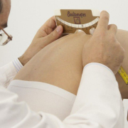
There is, of course, always a chance that these curves will become more pronounced as the youngster grows, even, in some cases, to the point of requiring the use of a brace. But it is also true that most of them will remain stable over time without reaching the minimum criteria for a diagnosis of scoliosis. Certain factors may possibly be associated with an increased risk of scoliosis progression: a positive family history of scoliosis, laxity of ligaments, flattening of physiological thoracic kyphosis, a greater than 10° angle of trunk rotation (ATR), and growth spurts. All these factors should be evaluated by the attending physician.
So, should we be treating these youngsters? In short, no. First of all, it is worth remembering, that the main aim of conservative treatment of scoliosis is to improve the patient’s appearance, but curves as mild as this rarely have an aesthetic impact; at most there may be some slight asymmetry of the trunk, but nothing that can be considered to exceed physiological parameters. With very rare exceptions, the only advice necessary in these cases is to opt for clinical monitoring of the patient, which can be considered to all intents and purposes a treatment, in the sense that it allows us to overcome the critical phases of development (which also correspond to the periods of greatest risk of progression of scoliosis) and also to intervene if any progression does occur. Monitoring is the first step in an active approach to idiopathic scoliosis, and it consists of clinical evaluations performed at regular intervals, ranging from every 2-3 months to every 36-60 months depending on the single case.
In conclusion, any active treatment in this population of patients is actually overtreatment. Even just specific exercises, whose prescription constitutes first therapeutic step after monitoring alone, would cost these youngsters in time and effort, as well as being an economic cost.
A further aspect, not to be underestimated, is the psychological impact: starting a treatment amounts to confirming that the individual has a disease that needs to be treated, and this can lead them to start thinking of themselves as “sick”.
Furthermore, even though an exercise programme is not a particularly arduous undertaking, starting a treatment when there is no real need for one could compromise the youngster’s collaboration and commitment should a treatment be needed later on. This is an important consideration, because if their scoliosis does progress as they grow, specific exercises, rather than being useful, could become crucial, in order to avoid bracing for example.
1 – 2016 SOSORT guidelines: orthopaedic and rehabilitation treatment of idiopathic scoliosis during growth
https://pubmed.ncbi.nlm.nih.gov/29435499/
2 – Radiographic Changes at the Coronal Plane in Early Scoliosis. Xiong, B., Sevastik, J. A., Hedlund, R., & Sevastik, B. (1994). Spine, 19(Supplement), 159–164. doi:10.1097/00007632-199401001-00008

