Reliability of rasterstereography as an alternative to X-rays in the diagnosis of scoliosis and its monitoring over time
To date, scoliosis continues to be diagnosed through clinical evaluation and confirmation by radiological examination. Monitoring its progression often entails frequent radiological assessments, which carry risks associated with radiological exposure, especially in growing patients.
Even though a low-dose radiation radiological investigation (using the EOS Imaging System, also offered to our patients) has recently been developed, it would still be an advantage to have at our disposal radiation-free methods for monitoring scoliosis and its progression.
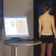
“Numerous systems that explore the topography of the posterior surface of the trunk (i.e., surface topography, ST) have been developed as alternatives to normal radiography, with the aim of reducing patient exposure to ionising radiation. And these systems can accurately reproduce the Cobb angle” explains ISICO physiatrist Carmelo Pulici. “One of them, Formetric rasterstereography, which involves surface detection and rasterstereography, has become an increasingly widespread radiation-free option in evaluating vertebral deformity. Thanks to structured light, this technique allows us to recreate an approximate 3D image of the spine’s shape”.
How does it work? The rasterographic projects parallel lines of white light onto the patient’s back.
The three- dimensional shape of the spine distorts these lines, producing a curved light pattern which is captured by a camera. A three-dimensional model of the back and spine is then constructed. ST “photographs” patients in their normal and habitual postures, avoiding some of the unnatural postural changes induced when they are positioned in front of an X-ray machine.
Unfortunately, however, research has failed to show a reliable correlation of diagnostic measurements between radiography (Cobb angle) and ST (1; 2).
“Cobb angle measurement is still the basis for the clinical treatment of scoliosis, and ST has not been shown to be an effective diagnostic substitute for traditional radiography”, Dr Pulici continues. “Specifically, recent studies have shown that ST does not meet the accuracy thresholds it needs to reach in order to be a useful primary diagnostic tool (it shows moderate accuracy) or a valid means of monitoring the progression of the curve (in this case its accuracy is low)”.
It is even less reliable in bracing therapy, where trunk remodelling means no longer a correspondence between what is observed from the outside and what is happening in the body.
Conversely, rasterstereography has demonstrated greater reliability in the measurement and monitoring, over time, of changes in the sagittal curves of the spine (lordosis and kyphosis). “We should also add that, as some research suggests, ST could be considered for use in early scoliosis screening of large populations (e.g., in schools)” Pulici says. In fact, it has been shown to be able to detect the presence of spinal deformity (but it is not accurate enough to quantify this) (2).
Is rasterstereography a valid noninvasive method for the screening of juvenile and adolescent idiopathic scoliosis? DOI: 10.1007/s00586-018-05876-0